Reading...
![]()
Play button
![]()
Play button
![]()
Use LEFT and RIGHT arrow keys to navigate between flashcards;
Use UP and DOWN arrow keys to flip the card;
H to show hint;
A reads text to speech;
79 Cards in this Set
- Front
- Back
|
What is the overall function of the auditory system? |
Transform acoustic information into mechanical activity and ultimately into electrochemical signals that are transmitted to the brain to provide us with what we call "hearing"
|
|
|
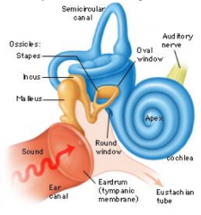
What are the sections of the auditory system?
|
- Outer ear
- Middle ear - Inner ear - Auditory nerve - Central auditory pathways |
|
|
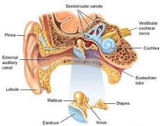
What are the components of the outer ear?
|
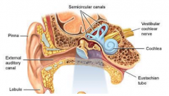
- Pinna
- External auditory meatus (canal) |
|
|
What are the components of the middle ear?
|

- Tympanic membrane
- Tympanic cavity - Ossicular chain (associated muscles, ligaments, and tendons; malleus, incus, stapes) - Eustacian tube |
|
|
What are the components of the inner ear?
|

- Oval window
- Cochlea - Vestibular structures |
|
|
What is the definition of sound?
|
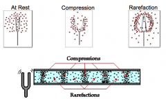
Longitudinal waves passing through a medium
|
|
|
What is the pinna? Function?
|
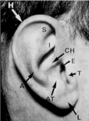
- Skin covered cartilaginous structure
- Channels and filters sound waves into the ear canal (Aka auricle) |
|
|
What can pits, tags, or other malformations of the pinna indicate?
|
Issues with ear development that may affect hearing
|
|
|
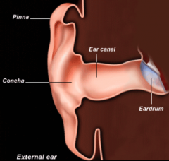
What are the boundaries of the external acoustic meatus (ear canal)?
|

- Concha (hollow area next to the ear canal)
- Eardrum |
|
|
What parts of the ear canal are cartilaginous? Bony?
|

- Outer 1/3 is cartilaginous (thick skin)
- Inner 2/3 is bony |
|
|
What is the function of the cartilaginous (outer 1/3) part of the ear canal?
|

- Skin is thick and contains wax and oil glands
- Wax/oil lubricate the canal and protect the ear from foreign objects and debris |
|
|
What can happen if you stimulate deep in the ear canal?
|
Cause referred sensation to vagus nerve, making people cough
|
|
|
Together, what are the benefits of the pinna and ear canal?
|
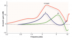
- Provide a boost in high frequency (~3000 Hz) sound intensity
- Canal (C) boosts from ~2000-3000 Hz - Pinna (P) boosts from ~5000-6000 Hz |
|
|
What is the role of the middle ear?
|

Helps to overcome impedance mistmatch between two media:
- Air in outer ear - Fluid in inner ear |
|
|
What are the ossicles?
|
- Malleus
- Incus - Stapes |
|
|
What is attached to the tympanic membrane? Function?
|

Malleus - vibrates in response to sound pressure waves funneled in by the external ear
|
|
|
What is attached to the oval window?
|

Footplate of the stapes
|
|
|
How does the middle ear overcome the impedance mismatch between the air medium (outer ear) and the fluid medium (inner ear)? What is the total gain of the middle ear?
|
Ossicles of middle ear amplify sound vibration by:
- Lever mechanism (~2 dB) - Area difference between tympanic membrane and footplate of stapes (increases force per unit area) (~23 dB) - Buckling of tympanic membrane (~6 dB) *Total gain ~31 dB |
|
|
Why is the size of the stapes footplate important?
|
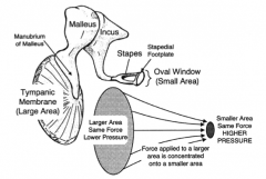
- Tympanic membrane is 14-15x bigger than stapes footplate
- Increases the force/unit area of the vibrations of the stapes footplate on the oval window of inner ear |
|
|
How much alone does the difference in area of the tympanic membrane to oval window increase the sound force?
|

~23 dB (of total ~31 dB for entire middle ear)
|
|
|
How does the lever action of the middle ear help with the impedance mismatch between air and fluid media? Gain?
|
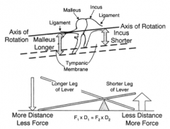
- Ossicular chain - length of malleus is longer than the long process of the incus
- Incus rotation has to cover less distance and is able to provide more force - Ratio: 1.3:1 - Gain of ~2 dB |
|
|
What is the shape and structure of the tympanic membrane?
|
- Cone-shaped
- 3 layer translucent membrane - ~1cm^2 vibratory surface |
|
|
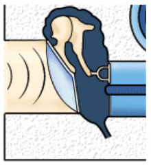
How does buckling of the tympanic membrane help to overcome the impedance mismatch of the air to fluid media?
|
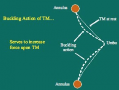
- Buckling of TM when it vibrates applies almost twice the force to the malleus
- Gain of ~6 dB |
|
|
How does the middle ear protect the ear when loud sounds are present?
|

- Protective feedback to dampen vibration of ossicles
- Tensor tympani m. (attached to malleus) - Stapedius m. (attached to neck of stapes) - When these contract they dampen the vibration of the ossicles |
|
|
What muscle attaches to the malleus? Innervation?
|

Tensor Tympani - Trigeminal n.
|
|
|
What muscle attaches to the neck of the stapes? Innervation?
|

Stapedius - Facial n.
|
|
|
What are the two functions of the inner ear?
|
- Hearing
= Balance |
|
|
What bony structure is the inner contained within?
|
Petrous apex of the temporal bone, encased by the osseous or bony labyrinth
|
|
|
What are the three sections of the bony labyrinth?
|

- Vestibule
- Cochlea - Semicircular canals |
|
|
What is the initial communication between the middle and inner ears?
|
- At the oval window of the vestibule where the stapes footplate abuts the oval window membrane
- At the basal end of the cochlea is the round window, which communicates with the middle ear |
|
|
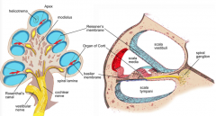
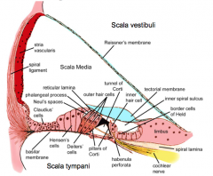
What are the components of the cochlea?
|

- Modiolus
- Osseous spiral lamina - Scala vestibuli - Scala tympani - Scala media - Helicotrema - Basilar Membrane - Reissner's Membrane |
|
|
What is the core of the cochlea?
|

Modiolus
|
|
|
What is the modiolus?
|

- Core of the cochlea
- Highly porous bone - Allows passage of auidtory nerve fibers |
|
|
How do the auditory nerve fibers get from the internal auditory meatus to the hair cell synapse?
|

Via the modiolus in the cochlea
|
|
|
What is the osseous spiral lamina?
|
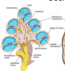
- Extension from the modiolus into the osseous labyrinthine space
- Bony shelf that coils around the center of the cochlea and provides partial division of upper and lower cochlear chambers - Also is the point of attachment for the basilar membrane |
|
|
What does the osseous spiral lamina partially separate?
|

Partially divides the upper and lower cochlear chambers into the scala vestibuli and scala tympani
|
|
|
What is the part of the cochlear labyrinth where the scala tymapni and scala vestibuli meet?
|
Helicotrema
|
|
|
What is the helicotrema?
|

- The part of the cochlear laybyrinth where the scala tympani and scala vestibuli meet
- At apex of the cochlea |
|
|
What does the basilar membrane attach to? What does it separate?
|
- Attaches to the osseous spiral lamina
- Separates the scala tympani and the scala media |
|
|
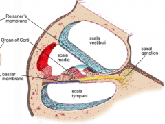
What are the three chambers of the cochlear labyrinthe? What separates them?
|

- Scala Vestibuli
- Reissner's Membrane - Scala Media - Basilar Membrane - Scala Tympani |
|
|
What is the sensory organ of hearing?
|

Organ of Corti
|
|
|
What is the stria vascularis? Where is it located?
|

- Highly vascular tissue that is responsible for metabolic environment of the scala media
- Along the lateral wall of the membranous labyrinth |
|
|
What supplies blood and nutrients to the scala media?
|

Stria vascularis (red area)
|
|
|
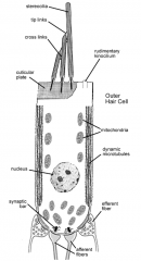
What are the components of the organ of Corti?
|

- Many types of epithelial cells and structures
- One row of inner hair cells - Three rows of outer hair cells - Supporting cells |
|
|
What kind of fluid is found within each of the three cochlear chambers?
|
- Scala vestibuli - perilymph
- Scala media - endolymph - Scala tympani - perilymph |
|
|
What are the characteristics of the perilymphatic fluid / perilymph?
|
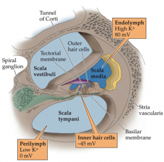
- Found in the scala vestibuli and scala tympani
- Contains high conc. of Na+ and low conc. of K+ - Similar to CSF and blood serum |
|
|
What are the characteristics of the endolymphatic fluid / endolymph?
|

- Found in the scala media
- Contains a high conc. of K+ and low conc. of Na+ - Similar to intracellular fluid |
|
|
How do the relative concentrations of Na+ and K+ differ between the perilymph and the endolymph?
|

- Perilymph - High Na+, Low K+
- Endolymph - Low Na+, High K+ |
|
|
What maintains the high K+/low Na+ within the endolymph?
|
Stria vascularis
|
|
|
How does the endolymphatic sac communicate with the membranous labyrinth?
|
- Endolymphatic duct
- Vestibular aqueduct |
|
|
What can happen if there are disorders of the endolymphatic system?
|
- Severe auditory and vestibular symptoms
- E.g., enlarged vestibular aqueduct may lead to sudden and progressive sensorineural hearing loss in children, particularly following head trauma |
|
|
What innervates the hair cells?
|

Dendrites of afferent bipolar neurons (cell bodies in spiral ganglion)
|
|
|
What percentage of the dendrites of afferent bipolar neurons contact inner hair cells? outer hair cells?
|
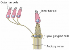
- 90-95% to inner hair cells (many afferent fibers synapse on the same inner hair cell)
- 5-10% to outer hair cells (single afferent fibers branch to synapse on several outer hair cells) |
|
|
Where do efferent fibers have their cell bodies? Where do they synapse?
|
- Cell bodies in superior olivary complex of brainstem
- Synapse directly on outer hair cells - Synapse on afferent fibers of inner hair cells |
|
|
What is the function of inner and outer hair cells?
|

Receptor cells that transduce mechanical movement into an electrochemical signal to stimulate the auditory nerve
|
|
|
Approximately how many inner hair cells are there? Outer hair cells?
|
- IHC ~3500
- OHC ~12,000 |
|
|
What are the characteristics of inner hair cells?
|

- Passive transducers in auditory system
- Highly metabolic |
|
|
What are the characteristics of outer hair cells?
|
- Contain microfilaments and microtubules along length of cell
- Allows for contractile behaviors - Motile activity results in increased basilar membrane motion |
|
|
What is the cochlear amplifier mechanism?
|
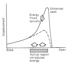
- Positive feedback within cochlea that provides acute sensitivity in the mammalian auditory system
* Outer hair cells increase basilar membrane deflection - Leads to increased amplitude and frequency selectivity of sound vibrations (dashed line represents basilar membrane alone; solid line represents with the positive feedback of OHC) |
|
|
What structures are on the apical portion of all hair cells?
|

- Thickened region - cuticular plate - forms reticular lamina
- Stereocilia - bundles of actin filaments |
|
|
What is rooted in the cuticular plate of each hair cell and projects through the reticular lamina?
|

Bundles of actin filaments called stereocilia
|
|
|
What are stereocilia?
|

- Bundles of actin filaments
- Stiff, hair-like structures that deflect with mechanical disturbance - Found on apical portion of all hair cells |
|
|
How are stereocilia connected? Why are they connected?
|
- Filamentous cross-links
- Tip-links - Allows the stereocilia to move as a unit when the longer stereocilia are deflected |
|
|
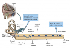
If you uncurled the basilar membrane how would its structure differ at the base vs. the apex? How does this determine the frequencies it responds to?
|
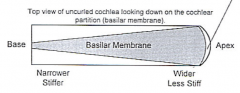
- Base: narrow/stiff - high frequencies
- Apex: wide/floppy - low frequencies |
|
|
What is the tonotopic organization of the basilar membrane?
|

- Base - high frequencies
- Apex - low frequencies |
|
|
What happens when pressure waves are transmitted from the middle ear to the cochlea?
|
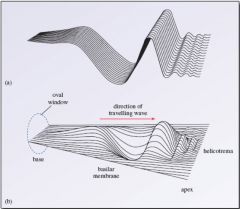
- Cochlear fluid is displaced
- Wave-like motions begin along length of basilar membrane - Forms a traveling wave |
|
|
Movement of what fluid causes deflection of the stereocilia?
|

Endolymph
|
|
|
What happens when stereocilia are deflected?
|
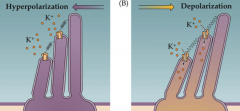
- Tiplinks open and close pores on top of stereocilia
- Positive ions (K+) from endolymph flow in and depolarize hair cells |
|
|
What happens when K+ flows into deflected stereocilia?
|
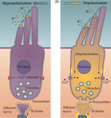
- Depolarization of hair cells
- Leads to Ca2+ flowing in - Vesicles release NT - Stimulates auditory nerve fibers to brain |
|
|
What causes the stereocilia to be deflected?
|
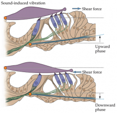
- Interaction of stereocilia with tectorial membrane (purple structure)
- Upward phase of movement of the basilar membrane |
|
|
Normal hearing should be able to detect what range of frequencies? What level of pitch?
|

- Entire range of frequencies (125 - 8000 Hz)
- All frequencies should be heard at low pitch (0-10 dB) |
|
|
What are common causes of hearing loss?
|
- Genetics
- Medications - Infections - Trauma (e.g., noise exposure) - Age (aka presbycusis) - Combination |
|
|
What are the main types of hearing loss?
|
- Conductive
- Sensorineural - Mixed |
|
|
What happens in conductive hearing loss?
|
- Occlusion of dysfunction of the external and/or middle ear
- On an audiogram, all frequencies will require a similar threshold |
|
|
What happens in sensorineural hearing loss?
|
- Dysfunction of the cochlea and/or auditory nerve
- Certain frequencies can be at a normal threshold while others will be at a higher threshold |
|
|
What happens in mixed hearing loss?
|
Sensorineural loss with a conductive component
|
|
|
How do you test hearing?
|
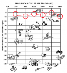
- Measuring detection thresholds to stimuli
- Plot detection thresholds on an audiogram |
|
|
What is the name for hearing loss due to old age? Which frequencies are particularly harder to hear?
|
Presbycusis (lose higher frequencies)
|
|
|
What are some therapies for hearing loss?
|
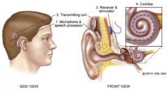
- Hearing aids - amplify sounds of specific frequencies (may not be enough)
- Cochlear implants (in picture) |