Reading...
![]()
Play button
![]()
Play button
![]()
Use LEFT and RIGHT arrow keys to navigate between flashcards;
Use UP and DOWN arrow keys to flip the card;
H to show hint;
A reads text to speech;
20 Cards in this Set
- Front
- Back
|
Def of hemianopia
|
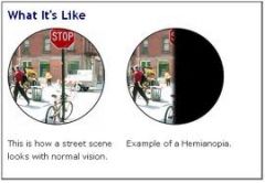
Loss of one half of the visual field
|
|
|
Heteronymous anopia
|
Different visual field defect in both eyes
|
|
|
Organization of the Visual Field:
• Def of point of fixation • How the extension of the visual field is measured • Other terms for the visual hemifields, specifically for the right eye • The generic names for the 4 quadrants of the visual field • Describing the position of the blind spot with respect to quadrants and point of fixation |
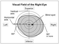
Organization of the Visual Field:
• The center of the visual field • It is measured in degrees of maximum deviation from the point of fixation • For the right eye - L visual hemifield = nasal visual hemifield - R visual hemifield = temporal visual hemifield • Superior (left and right), Inferior left and right) • It is ~15 degrees to the right of the point of fixation |
|
|
The visual field and the retina:
• Describe how the visual field is projected onto the retina |
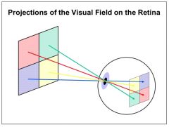
The visual field and the retina:
• It is projected inversely on to the retina - The superior half is projected onto the inferior half - The nasal visual hemifield is projected onto the temporal hemiretina |
|
|
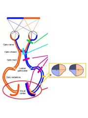
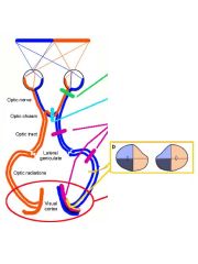
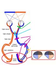
Visual Pathway 1: The Left Superior Quadrant
• The location of its retinal targets (2) • The course of the above retinal fibers and where they synapse • After the synapse, - the name of these new fibers - the pathways they use and their synapse > the relative location of this synaptic target |
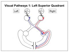
Visual Pathway 1: The Left Superior Quadrant
• Its retinal targets - The inferior portion of the nasal hemiretina of the left eye - The inferior portion of the temporal hemiretina of the right eye • The course after the retina - The left inferior nasal hemiretina crosses the optic chiasm - The right inferior temporal hemiretina stay ipsilateral * Both will synapse at the right LGN • After the LGN, - the fibers emerge as the optic radiation with the > contralateral fiber using the nasal radiation pathway and > ipsilateral fiber using the temporal radiation pathway - Both will synapse in the inferior portion of V1 > below the calcarine sulcus |
|
|
Visual Pathway 2: The Left Inferior Quadrant
• The location of its retinal targets (2) • The course of the above retinal fibers and where they synapse • After the synapse, - the name of these new fibers - the pathways they use and their synapse > the relative location of this synaptic target |
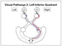
Visual Pathway 2: The Left Inferior Quadrant
• Its retinal targets - The Superior portion of the nasal hemiretina of the left eye - The Superior portion of the temporal hemiretina of the right eye • The course after the retina - The left Superior nasal hemiretina crosses the optic chiasm - The right Superior temporal hemiretina stay ipsilateral * Both will synapse at the right LGN • After the LGN, - the fibers emerge as the optic radiation with the > contralateral fiber using the nasal radiation pathway and > ipsilateral fiber using the temporal radiation pathway - Both will synapse in the superior portion of V1 > above the calcarine sulcus |
|
|
The Primary Visual Cortex
• Location of Brodmann's area • Where the major portion of V1 is represented > Side of the brain > describe w/ respect to a sulcus • its blood supply (branches of major source, major source and minor source) |
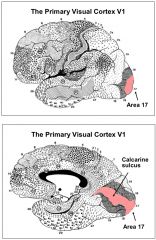
The Primary Visual Cortex
• 17 • The major portion of V1 is represented > on the medial side of the brain > along the banks of two gyri, superior & inferior to the calcarine sulcus • its blood supply - Major: Calcarine branches from the PCA - Minor: MCA |
|
|
Organization of V1:
• Term used for the topographic organization present here and what it means • Where the following halves of the visual field are represented - left half - right half - inferior portions - superior portions - center > also, another name for this portion of the visual field - peripheral |
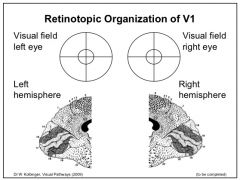
Organization of V1:
• Retinotopic - refers to positions of stimuli on the retina • Where the following halves of the visual field are represented on V1 - left half --> right hemispheric V1 - right half --> left hemisheric V1 - inferior portions --> superior to calcarine sulcus - superior portions --> inferior to calcarine sulcus - center --> close to the occipital pole > another name is the macular region - peripheral --> closer to the parieto-occipital sulcus |
|
|
Two types of columns in V1
|
Ocular dominance and Orientation columns
|
|
|
Describe the wiring of Ocular dominance columns from sensory fibers of the LGN
|
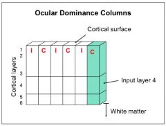
• Sensory fibers from one eye of the LGN do not terminate in the same area of V1 as that of the contralateral eye
• Separate areas/columns in the cortex exist as dedicated inputs to each eye |
|
|
Discuss the type of experiment performed by Hubel and Wiesel that lead to the dscovery of orientation columns.
- Include the general name of the experiment, the region of the brain and parameters measured and what was proved |
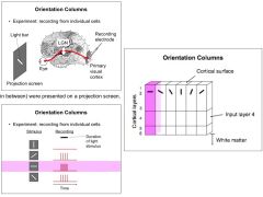
Experiment w/ single cell recording
• Using a recording electrode placed in V1, the activity of single cells were recorded by presenting the subject with different orientations of horizontal bars of light • Cells that showed the most sensitivity to a particular orientation had the highest frequency of APs recorded • The results concluded that: - V1 was structured into columns, in this instance for orientation - every cell in one cortical column has a preference for a certain orientation of a light bar. - the preferred orientation in neighboring orientation columns is different |
|
|
Two specialized parallel pathways for visual information, where their cells originate, where they synapse, the type of stimuli they discriminate and their trajectory after synapsing (this trajectory has 3 names)
|
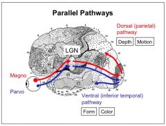
Magnocellular Pathway
- consist of M cells in the retina that synapse in the 1st two layers of the LGN - They discriminate for motion and depth - After the LGN, they form a dorsal/parietal pathway (what pathway) Parvocellular cells - consist of P cells in the retina that synapse in the last 4 layers of the LGN - They discriminate for form and color - After the LGN, they form a ventral/inferior temporal pathway (where pathway) |
|
|
The Confrontation Visual Test
• what it's used • how it's tested |
The Confrontation Visual Test
• used to test loss of vision • each quadrant of the visual field of each eye is tested - Patient and examiner are standing at twice the arm's length in front of one another - patient occludes one of their eyes - examiner moves their hand from the periphery to the center of the visual field to determine where it is seen first |
|
|
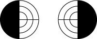
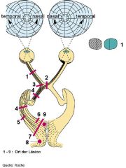
Type of lesion: Optic nerve
Visual defect produced: Possible origin: |
Type of lesion:
Visual defect produced: Monocular blindness Possible origin: Optic neuritis |
|
|
Type of lesion: Optic chiasm
Visual defect produced: Possible origin: |
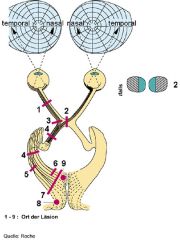
Type of lesion:
Visual defect produced: Bitemporal hemiopia Possible origin: Pituitary tumor |
|
|
Type of lesion: Optic tract
Visual defect produced: Possible origin: |
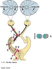
Type of lesion:
Visual defect produced: Homonymous hemianopia Possible origin: temporal lobe tumor |
|
|
Type of lesion: Temporal radiation (Meyer's Loop)
Visual defect produced: Possible origin: |
Type of lesion:
Visual defect produced: Homonymous Superior quadratic anopia Possible origin: Temporal or Occipital lobe tumor |
|
|
Type of lesion: Parietal radiation
Visual defect produced: Possible origin: |
Type of lesion:
Visual defect produced: Homonymous inferior quadrantic anopia Possible origin: Parietal or occipital lobe tumor |
|
|
Type of lesion: Visual cortex
Visual defect produced: Possible origin: |
Type of lesion:
Visual defect produced: Homonymous hemianopia Possible origin: Posterior cerebral artery dysfxn |
|
|
Macula sparing:
• definition • Usual etiology |
Macula sparing:
• visual field deficits not involving the macula • The MCA, the vessel supplying the occipital pole and macular vision, is not compromised but the PCA or any of its branches are |