Reading...
![]()
Play button
![]()
Play button
![]()
Use LEFT and RIGHT arrow keys to navigate between flashcards;
Use UP and DOWN arrow keys to flip the card;
H to show hint;
A reads text to speech;
12 Cards in this Set
- Front
- Back
- 3rd side (hint)
|
areas to observe on optic disc
|
1) shape- circular
2)color - normal yellow orange 3)borders- crescent? 4)optic cup - NORMALLY 40% of disc 5) blood vessels - measured in disc diameter Quadrants - inferior and superior temporal and Nasal |
|
|
|
Vessels - arteries be veins
|
1) arteries exit on nasal side, 2) are typically brighter red, 3) veins are larger 2:3, AV ratio decreases with hypertension 4) vessel walls visible - hypertension, normal age. 5) silver color = severe compromised blood flow
|
|
|
|
AV nicking
|
Narrowing of vein on either side of the arteriole. Gunns sign
|
|
|
|
AV RATIO
|
2:3, follow out 1-2 disc diameters DD
|
|
|
|
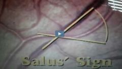
Salus' sign
|
|
|
|
|
Tortuosity
|
Sign of impending venous occlusion. 1:3 ratio.
|
|
|
|
Eye exam sequence
|
Optic disc, vessels, background, macula
|
|
|
|
Retinal hemorrhages
|
Vitreous, preretinal, intraretinal, subretinal
|
|
|
|
Most common hemorrhages
|
Intraretinal layer - 1) flame shaped 2) most common in diabetes and hypertension
|
|
|
|
Blot hemorrhages
|
Occur in ischemic areas closer to the temporal side common in DM.
|
|
|
|
Subhyloid hemorrhage
|
Blot appearance, causes blindness if over macula. Causes red haze if infiltrated in vitreous.
|
|
|
|
Exudates
|
Cotton wool patches
Common in the, diabetes, lupus, and aids. |
|