![]()
![]()
![]()
Use LEFT and RIGHT arrow keys to navigate between flashcards;
Use UP and DOWN arrow keys to flip the card;
H to show hint;
A reads text to speech;
68 Cards in this Set
- Front
- Back
|
5yo M with cavus feet that began to develop at 3yo. Decreased distal sensation to pinprick, decreased vibratory sensation, absent Achilles reflexes. Gower's sign negative. NCV delayed. Dx? |
Charcot Marie Tooth disease |
|
|
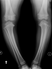
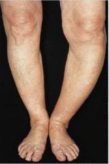
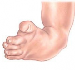
15 month old M presents to office for evaluation. Child began to walk at 9 months old. Dx? (pic of bow-legged child) |
physiologic genu varum (more common at this age opposed to Blount's) |
|
|
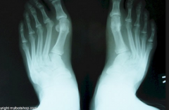
7 month old F feet turn in. bilateral moderately severe met adductus deformity. Initial recommended treatment?
|
serial casting |
|
|
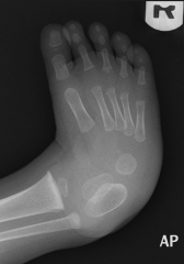
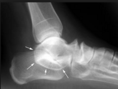
10yo F with acute pain posterior to medial malleolus and along medial aspect of midfoot. pain elicited with passive abduction of the forefoot. (pic of x-ray showing gorilloid navicular with small accessory navicular) Dx? |
posterior tibial tendinitis associated with an accessory navicular
|
|
|
Tx for the 10yo described in question 4 might include? (posterior tibial tendinitis associated with accessory navicular) |
Unna boot, anti-inflammatories, and orthotic therapy |
|
|
3yo F with spastic diplegic cerebral palsy with marked ankle equinus gait. no contracture at hamstring or hip level. passively 10º of ankle dorsiflexion available with knee extended. appropriate Tx? |
AFO with plantarflexion stop in conjunction with physical therapy |
|
|
12yo M suffers avulsion fracture of 5th met base during a basketball game. There's a separation between the fragments. Tx? |
immobilization in a BK cast |
|
|
12yo F with progressive unilateral left cavus foot deformity that began 2 years ago. distal motor and sensory deficit of the left foot is noted. Dx? |
spinal cord tumor (or tethered cord) -- always think these 2 with unilateral cavus foot |
|
|
12yo M returns for continued treatment of plantar verruca. Creamy exudate noted upon debridement. increased redness and swelling noted around the lesion. increased tenderness. Tx? |
culture of the exudate and antibiotic therapy |
|
|
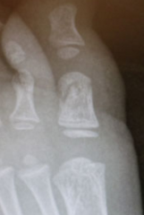
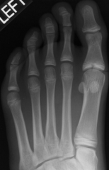
12yo M presents with painful hallux. Pt states he has been treated for recurrent 'ingrown toenails' in the past. nail is slightly elevated. the most important step in resolving the problem? (x-ray shows bony outgrowth of distal phalanx) |
excision of the exostosis |
|
|
which joint is most affected in juvenile rheumatoid arthritis? |
knee |
|
|
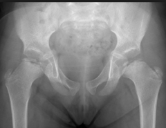
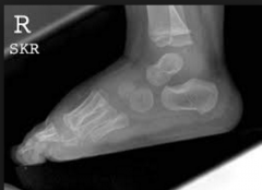
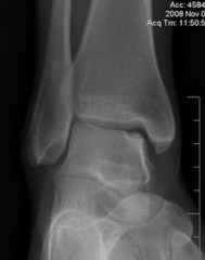
2yo breast-fed M presents with joint swellings of wrists, knees, and ankles. he does not take vitamins or drink milk. waddling gait noted. radiographs of the pelvis and ankle are shown. initial evaluation should include (what test to order)? |
serum calcium, phosphorus, and alkaline phosphatase analysis |
|
|
11 month old F presents with a flexible calcaneovalgus foot posture. her milestones are delayed and she slips through your arms when held under the axilla (vertical suspension sweet). these findings are consistent with? |
hypotonia |
|
|
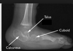
10yo M basketball player has right heel pain for 3 weeks. pain occurs after running and later at night. tenderness upon palpation of medial aspect of right calcaneus. radiographs reveal irregularity of the apophysis. Dx? |
calcaneal apophysitis |
|
|
12yo obese M presents with right groin and knee pain of 8 months duration. mother notes a right-sided limp and a right flat foot. No axillary hair. right leg is shorter, and internal rotation is limited on right side. Dx? |
SCFE - slipped capital femoral epiphysis |
|
|
12yo F presents with left sinus tarsi pain. left STJ ROM is restricted compared to right side. Dx? |
subtalar coalition |
|
|
6 month old F presents with a right 'up and out' deformity present since birth. plantarflexion of the ankle and supination of the foot is restricted. Forefoot varus of 30º noted. Deformity is? |
congenital calcaneovalgus |
|
|
3yo M presents because of his parents concerned of his legs when he walks. what could be a potentially significant finding? (pick one) -15º outtoe B/L -hip rotation of 30º internal rotation and 60º external rotation -genu varum -Bleck's test at the 2nd digit B/L |
genu varum -- should be knock-knee by this age |
|
|
5 month old F presents with left TEV deformity that has been casted since birth. recent x-rays show signs of a developing rocker bottom deformity. physician should consider? |
posterior medial release at 6-8 months of age |
|
|
5yo M with clumsy gait and difficulty rising from the floor. problem started 1 year ago. Pes planovalgus deformity noted as well as a tight heel cord. Dx? (pic shows a positive Gower's sign) |
muscular dystrophy |
|
|
6yo F presents with pruritic scaling of the plantar surface of the feet. Personal and Family Hx of asthma. an extra skin crease is noted under the eyes. Dx? |
atopic dermatitis |
|
|
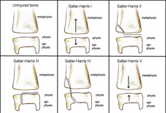
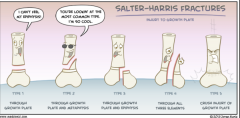
radiographic pictured? (just gonna put a pic of all types, answer was salter harris type 3) |
|
|
|
6to F presents with ligamentous laxity and long, narrow, severely pronated feet. she is in 99th percentile for height. arm span is longer than her total height and echocardiogram shows aortic dilatation. Dx? |
Marfan syndrome |
|
radiographs pictured here are consistent with? (google imaged this) |
TEV |
|
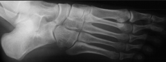
radiograph pictured is? (google imaged this) |
Calcaneonavicular bar |
|
3yo M presents for evaluation of persistent intoe gait and 'bowed legs'. radiographs taken at 13 months and 25 months old were read as within physiological normal limits. exam reveals severe internal tibial torsion, bowing of the legs, and a lateral knee thrust during ambulation. radiographs ordered again. Clinical impression? (google images) |
Blount's disease |
|
|
4yo M presents with left-sided toe walking and a painful left heel that began 2 weeks ago. a small, erythematous lesion noted on the plantar surface of left heel. no portal of entry noted. radiographs are ordered. next step? |
recommendation for surgical consultation (idk what the pic was) |
|
|
44 pound, 6yo M develops a mild soft tissue infection of the left plantar heel area while undergoing treatment for a plantar verruca. culture of the exudate indicates sensitivity to Keflex. Recommended 25-50mg/kg per day dose given every 12 hours for pediatrics (oral suspension). write the Rx. |
Keflex Oral Suspension 250mg/5mL DTD 100mL Sig.: 1tsp. every twelve hours for 10 days |
|
|
55 pound M prescribed Augmentin Oral Suspension. Recommended 25mg/kg per day. Comes in 200mg/5mL or 400mg/5mL. Write the Rx |
Augmentin Oral Suspension 200mg/5mL DTD 150mL Sig.: 1.5 tsp. every 12 hours for 10 days |
|
|
3.5yo F presents with asymmetric genu valgum deformity and LLD (left 1/4" shorter than right side). Differential Dx includes? |
-coxa vara -intrinsic bone disease (rickets, renal osteodystrophy) -abnormal epiphyseal plate growth due to trauma or infection -answer = all of the above |
|
|
5yo M presents with pain over the dorsomedial aspect of the right midfoot. STJ and MTJ ROM are unrestricted B/L. Gait analysis reveals excessive supination of the right foot during the stance phase and a right-sided antalgic limp. Radiographs are shown(silver dollar sign of navicular is seen). Dx? |
Kohler's disease |
|
|
5yo F with symmetrical, non-pruritic rash on weight-bearing surface of ball area of forefoot. Skin on the ball area and weight-bearing plantar aspect of digits has a shiny glazed appearance with some fissuring. the interdigital web spaces are unaffected. the condition is worse in the winter and improves in the summer months. Dx? |
Juvenile plantar dermatosis |
|
|
13yo F with symmetrical patches of red, scaly skin on the extensor surfaces of her legs and arms. the scaling is silver colored and dry. nail changes associated with this disorder? (give 3 answers) |
-subungual keratoses and pitting of the nails -splinter hemorrhages -onycholysis (psoriatic nail changes) |
|
|
14yo F runner complains of pain and tingling of the anterior lateral aspect of the left tibia and numbness of the left foot when running. the pain has been intermittent for the last 7 months. patient runs about 15-20 miles per week. neurologic exam reveals decreased sharp-dull sensation of the proximal lateral aspect of the left tibia. light touch sensation of the left foot is also reduced. Dx? |
compartment compression syndrome -- exercise induced |
|
|
1yo F presents with dystrophic hallux nails and absent thumb nails. Ultrasound reveals absent patellar growth centers. Dx? |
nail patella syndrome |
|
what congenital foot deformity is shown on this lateral radiograph? (google images) |
vertical talus -- severely plantarflexed, very rigid |
|
what orthopedic deformity is evident in this young patient? (google images) |
genu varum -- big gap between the knees by definition is genu varum even though it may appear as tibial varum |
|
13yo F presents with pain of the proximal phalanx of the left hallux. 2 weeks earlier she had tried to block a falling desk with her foot. Radiograph indicates? (best pic I could find online) |
comminuted intra-articular fracture (picture may actually be an extra-articular fracture) (nonetheless, other choices of Salter Harris type fractures are wrong because no epiphysis is involved) |
|
|
11yo F presents with a painful, swollen right ankle. Pt experiences post-static dyskinesia and complains of right medial ankle sprains. swelling of the left medial ankle is noted. pain is elicited upon palpation of the areas under the medial and lateral malleoli. radiographs indicate medial soft tissue swelling of the right ankle, a lowered calcaneal inclination angle and increased talar declination. Rheumatoid blood profile indicates an elevated ESR and ANA. Slit lamp exam reveals inflammation of the anterior chamber of the eye. Dx? |
pauciarticular rheumatoid arthritis |
|
|
6yo M presents to office for toe-walking gait that began with ambulation at 9 months of age. He toewalks 100% of the time. Exam reveals hyperreflexia of the lower extremities. Babinski response is extensor B/L and ankle clonus is noted. Good proximal and distal motor strength noted. Dx? |
spastic diplegia |
|
|
What would be the most likely next step in the evaluation of the patient described in question 41? (spastic diplegia patient) |
Brain MRI -- not EMG/NCV which is for peripheral neuropathy |
|
|
6yo spastic diplegic demonstrates negative 40º AJ dorsiflexion B/L with knee flexed and negative 50º AJ dorsiflexion with knee extended B/L. Hip ROM is 40º lateral and 50º medial rotation with hip extended. He is unable to tolerate an AFO. Next step in management? |
evaluate for bilateral tendo Achilles lengthenins (too much contracture for an AFO) |
|
|
6yo F born with bilateral dislocated hips, convex pes valgus of the feet, and flexion deformities of the wrists and fingers. Atrophy of the limbs and fixed extension of the knees was also noted at birth. Hx is consistent with? |
arthrogryposis multiplex congenita |
|
|
13yo F presents with subungual and periungual firm skin colored nodules of the halluces and fingers. 'Acne-like' rash appears on the face. Hx of seizures noted. Dx? |
tuberous sclerosis |
|
|
5yo F with a low-grade fever and sore throat presents with fluid filled vesicles of the hands and feet. Lesions are painless, non-pruritic, and not centrally umbilicated. Erosions of the upper palate noted. No lesions of the trunk evident. Hx consistent with? |
coxsackie virus 16 -- Hand-foot-mouth disease |
|
|
17yo M developed mildly pruritic bullae of the dorsum of the hands and feet while incarcerated in a juvenile detention center. after several days the bullae ruptured causing a yellowish crusting over a centrally ulcerated area. Dx? |
impetigo -- yellowish crusting is the giveaway |
|
|
16yo F presents with painful, red nodules of the anterior aspect of the tibiae measuring 4-6cm. Vascular exam WNL. PPD test positive. Hx suggests? |
erythema nodosum as a manifestation of tuberculosis |
|
|
6yo F presents with complaint of pain behind the right knee. pain occurs while standing barefoot for long periods and after tae kwon do classes. Knee feels better when she is wearing running shoes. Problem started 4 months ago. A firm, well-circumscribed soft tissue mass noted behind the right knee. More prominent with knee extension and less with knee flexion. Medical Hx unremarkable. Dx and next step in management of the patient? |
Baker's cyst -- referral to orthopedist |
|
|
16yo M adolescent presents with a pigmented lesion of the dorsum of the right ankle. Mother and grandmother have the same birthmark. Lesion has been present since birth but smaller pigmented areas have developed around the edges and the surface has become irregular. A dark bluish tint is noted and the pigmentation is uneven. Clinical impression pending biopsy results? |
malignant blue nevus |
|
9yo M presents with severe right groin pain and a right sided limp. The right leg is shorter and hip abduction is limited on that side. Hip pathology is suspected and the patient is promptly referred to the orthopedist. Hip radiographs taken. Dx? (google images) |
Legg-Calve-Perthe's disease |
|
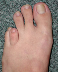
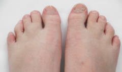
12yo F presents for evaluation of 'short toes'. Clinical picture is consistent with? |
brachymetatarsia |
|
|
The condition seen in question 51 is associated with? (brachymetatarsia patient) (3 answers) |
-pseudohypoparathyroidism -Down syndrome -Turner's syndrome |
|
|
16yo healthy M presents with 'painful blisters' on the bottom of his feet of 3 weeks duration. The ball and toe pad areas of the feet appear as if they have been immersed in water for a long period of time. Multiple pits are noted on the plantar aspect of the feet. Some of the pits have a black pigmentation that cannot be washed away. the feet are hyperhydrotic, bromhydrotic, and tender to palpation. the condition resolved dramatically with use of oral erythromycin and betadine soaks. Dx? |
Pitted keratolysis |
|
4yo M presents for evaluation of a partial fusion of the 2nd and 3rd digits of the feet. Parents would like a second opinion regarding possible treatment. The child has no discomfort or difficulty fitting shoegear. Dx and Tx? (google images) |
partial syndactyly -- no surgical intervention |
|
|
13yo M presents with a painful, chronic ulcer of the right lateral ankle. Past medical Hx includes rickets and sickle cell disease. No other medical problems. Most likely cause of the ulcer? |
sickle cell disease |
|
|
14yo M presents with redness and itching of the plantar aspects of the feet and interdigital spaces. Feet are hyperhydrotic. painful blisters noted on the plantar and lateral aspects of both feet. Tx? |
"popping" large, painful blisters with a sterile needle, betadine soaks, and topical spectazole (vesciulobullous tinea pedis) |
|
|
12yo F, her mother, and 9yo sister present with plantar calluses. younger sister also has calluses on her hands and elbows. Grandmother and great grandfather also have a history of plantar calluses. Dx? |
hereditary palmar plantar keratoderma |
|
13yo F presents with left lateral submalleolar pain during long periods of walking. Patient has bilateral residual clubfeet and underwent a posterior medial release at 11 months of age. X-rays indicate? (google images) |
Left talocalcaneal coalition |
|
|
10yo F figure skater presents with complaint of painful bumps on the back of her heels. pain is worse in ice skates but she is also experiencing pain in her sneakers. Tx options? |
-flare counter of the skate -U pad and Ipos silastic sock -custom orthoses in an extra depth ice skate (could be Haglund's deformity) |
|
What foot deformity is consistent with these DP and lateral radiographs? (google images) |
plantarflexed talus |
|
7yo African American F presents for evaluation of intoe gait. what deformity does she demonstrate? |
metatarsus adductus |
|
|
16yo 160lb. M presents with bilateral paronychias of the medial border of the hallux. What is the maximum dosage of 2% xylocaine that can be given to this patient? |
15mL |
|
|
9yo African American M presents with pain on the dorsum of the right foot. What bone is prominent of x-ray? (cant find a pic) |
middle cuneiform |
|
The clinical presentation of this 1 month old are consistent with? |
TEV |
|
17yo F presents with aching pain over the dorsal ankle. pain occurs 5-6 miles into the run. pain began 4 months earlier. she runs 12-15 miles a week. No Hx of trauma. Radiograph indicates? |
osteochondritis dissecans of talus |
|
|
Place the following lateral radiographs in correct sequence |
be able to identify TEV, plantarflexed talus, and vertical talus on x-ray |
|
17yo M presents with pain of the left hallux of 3 days duration. patient dropped his electric guitar on top of his left foot 3 days earlier and states the pain is 9/10 on VAS. Edema of left hallux is evident and pain is most acute upon palpation of the proximal phalanx of the left hallux. Radiograph shows? |
Salter Harris 3 |
|
|
10yo 105lb F presents with resistant multiple verrucae of the right foot. Use of cimetidine, Aldara, and 70% topical salicylic acid have been unsuccessful. Recent studies indicate some success with use of zinc sulfate (10mg/kg per day up to maximum 600mg) in children. Write a Rx for zinc sulfate tablets for 60 days. (Comes in 10mg, 30mg, 50mg, 225mg tablets). Given twice a day. |
Zinc Sulfate 225mg Disp 120 tablets Sig.: 1tab PO BID |