![]()
![]()
![]()
Use LEFT and RIGHT arrow keys to navigate between flashcards;
Use UP and DOWN arrow keys to flip the card;
H to show hint;
A reads text to speech;
83 Cards in this Set
- Front
- Back
|
Electrical Stimulation of the Brain (ESB) |
Stimulation technique. Electrical current delivered through electrodes directly contacting specific brain areas |
|
|
Electroencephalograph (EEG) |
Electrical Recording. Invented in 1929 by psychiatrist Hans Berger; Electrodes attached to scalp record electrical activity of groups of neurons (reading of brain activity by putting sensors on scalp) |
|
|
Computerized Axial Tomography (CT) |
Brain Imaging. Computer-enhanced X-ray of brain structure; takes pictures of narrow slice of brain (just looks at brain structure) |
|
|
Positron Emission Tomography (PET) |
Brain Imaging. Measures brain activity (metabolism, blood flow, and neurotransmitter activity); radioactive glucose injected into blood stream (activated areas of the brain become visible) |
|
|
Magnetic Resonance Imaging (MRI) |
Brain Imaging. Produces 3-D, high resolution pictures of brain mainly of structure; measures body's response to magnetic impulse; takes images several minutes apart |
|
|
Functional Magnetic Resonance Imaging (fMRI) |
Brain Imaging. Produces 3-D, high resolution pictures of brain mainly of structure; measures body's response to magnetic impulse; takes images only seconds apart; shows changes in brain activity |
|
|
Hemispherectomy |
Surgical procedure in which an entire hemisphere of the brain is removed |
|
|
Brain Plasticity |
Brain can change shape; new neurons are formed, taking functions of dysfunctional/impaired side of the brain (rewiring) |
|
|
Rasmussen Syndrome |
Seizures that cannot be controlled with medicine |
|
|
Split-brain Surgery |
Cutting the corpus callosum (connection between brain hemispheres); done on ''Joe'' to stop seizures |
|
|
Pre-frontal Lobotomy |
Cutting connections between frontal lobe and the rest of the brain; vegetative state |
|
|
Central Nervous System (CNS) |
Composed of the brain and the spinal cord |
|
|
Peripheral Nervous System |
Connects Central Nervous System (CNS) with muscles, glands, and sensory receptors |
|
|
Somatic Nervous System |
Subdivision of the Peripheral Nervous System; triggers voluntary muscle activation through motor neurons/nerves (efferent) |
|
|
Autonomic Nervous System |
Subdivision of the Peripheral Nervous System; Gland and involuntary muscle activation; Sympathetic (fight-or-flight; activated mode) and Parasympathetic (deactivation) |
|
|
Sympathetic Autonomic Nervous System |
Mobilize bodily resources for emergency (fight-or-flight); i.e. drains blood from periphery of body to reduce bleeding in injury, slows down digestion, triggers adrenal glands (adrenaline) |
|
|
Parasympathetic Autonomic Nervous System |
Conserves bodily resources (i.e. save and store energy, slow heart rate, decrease blood pressure, promote digestion) |
|
|
Cerebrospinal fluid (CSF) |
Nourishes brain; serves as protective cushion |
|
|
Glia |
Structural support of neuron, insulation, and nourishment |
|
|
Neuron |
Soma (body), dendrites (receive info), axon (includes myelin sheath + terminal buttons; transmits info through neurotransmitters |
|
|
Myelination |
Allows for more accurate information transmissions |
|
|
Properties of Axon |
Contains electrically charged particles called ions (Na+, K+, Cl-, A-); Cell membrane acts as a selective filter, at different times allowing different ions through; stimulation from surrounding neurons sets in motion the opening and closing of certain ion channels along axon of a neuron. |
|
|
Resting Neuron |
Higher concentration of negative ions inside cell; Action potential = ~ -70mV; Polarization |
|
|
Active Neuron |
Stimulation of neuron results in higher concentration of positive ions inside cell; action potential = ~ +40 mV; Depolarization |
|
|
Action Potential |
the first causes a chain effect (multiple action potentials); exchange of chemicals through the axon; leads to transmission of information to the next cell |
|
|
Depolarization requires? |
Sufficiently strong stimulation coming in from other neurons; insufficient graded potentials won't start an action potential |
|
|
All-or-none Law |
Neuron fires at full-strength or not at all (starting action potential and nervous impulse or not) |
|
|
Absolute Refractory Period |
When neuron can't fire, not sufficient to start action potential |
|
|
Relative Refractory Period |
When neuron fires at elevated threshold; starting action potential |
|
|
Synaptic Transmission |
Following action potential, neurotransmitters are released from synaptic vesicles, travelling across the synaptic cleft and finally binding to 'custom' receptor sites in postsynaptic neuron, following, they activate or inhibit the postsynaptic neuron; after this process, neurotransmitters are either broken down or reabsorbed (reuptake) by cell to be reused |
|
|
Neurotransmitters |
Chemicals that transmit information from one neuron to another; send two types of messages: excitatory (postsynaptic potential that increases probability of action potential in postsynaptic neuron) or inhibitory (postsynaptic potential that decreases probability of action potential in postsynaptic neuron); linked to specific behavioural phenomenon; react to psychoactive drugs |
|
|
Acetylcholine (ACh) |
Activates voluntary muscles; curare (paralytic drug) is an antagonist; contributes to attention, arousal, memory; undersupply related to memory loss (Alzheimer's); memory drugs are agonists; Nicotine is an agonist |
|
|
Dopamine (DA) |
Monoamine; Contributes to voluntary movement and positive emotional states; undersupply linked to Parkinson's disease; L-DOPA, cocaine, and amphetamine are agonists; Oversupply linked to Schizophrenia; some anti-psychotic drugs are antagonists; marijuana-schizophrenia link identified |
|
|
Serotonin |
Monoamine; involved in regulation of sleep, wakefulness, eating behaviour, and aggression; imbalances linked to psychological disorders such as depression; Prozac works by preveting the reuptake; eating disorders; obsessive-compulsive disorders; ecstacy acts as agonist |
|
|
Norepinephrine |
Monoamine; low levels linked to depression; amphetamines and cocaine are agonists |
|
|
Gamma-aminobutyric Acid (GABA) |
Amino acid; Produces inhibitory postsynaptic potential; imbalances linked to anxiety disorders |
|
|
Glutamate |
Amino acid; contributes to learning and memory |
|
|
Endorphins |
Opiate-like substances that act as pain modulators; morphine and heroin are agonists; explain runner's high |
|
|
Three Major areas of Brain |
Hindbrain, Midbrain, Forebrain |
|
|
Hindbrain |
Brainstem: Medulla (heart rate, blood circulation, respiration, muscle tone, reflexes), and Pons (sleep, dreaming, arousal, connects brain stem to cerebellum).
Cerebellum: Coordination of movement, equilibrium, balance, neural circuits run from cerebellum to pre-frontal cortex (attention, planning, visual perception) |
|
|
Midbrain |
Segment of brainstem between hindbrain and forebrain; important in integrating sensory processes; seat of dopamine producing neurons that project to higher cerebral centres; reticular formation runs from hindbrain to midbrain and beyond (modulates muscle reflexes, breathing, pain, perception; regulates sleep and arousal) |
|
|
Forebrain/Cerebrum |
Thalamus: reception and redirection of signals to appropriate brain area
Hypothalamus: controls ANS, endocrine system connection (hormones), regulates survival drives (fighting, fleeing, feeding, mating)
Limbic System: regulation of emotion, memory, motivation (includes thalamus, hypothalamus, hippocampus [formation and retrieval of memory], and amygdala [aggression, fear, pleasure]) |
|
|
Frontal Lobe |
Speech - Broca's area (left hemisphere); motor cortex; prefrontal cortex (calm decision-making; problem results in lack of control of emotions and impulsion) |
|
|
Parietal Lobe |
Somatosensory cortex (sense of touch); spatial sense and navigation |
|
|
Temporal Lobe |
Speech - Wernicke's area (left hemisphere); auditory cortex |
|
|
Laterization |
Localization of some functions in one of the two brain hemispheres |
|
|
Left Hemisphere |
Verbal processing (Broca's and Wernicke's areas); logical and mathematical abilities; positive emotions; right visual field processed |
|
|
Right Hemisphere |
Holistic hemisphere; spatial relation (sense of direction); melodies (musical abilities); negative emotions; left visual field processed |
|
|
Information Processing |
We look with our eyes, yet we see with our brain; seeing is more than the vision of the environment, it's the processing of information |
|
|
Sensation |
Stimulus-detection process by which sense organs respond to and translate environmental stimuli into nervous impulses that are sent to the brain |
|
|
Perception |
Active process of organizing this stimulus input and giving it meaning |
|
|
Visual Sensory System |
stimulated by light waves; rods and cones are receptors located in the retina of the eye |
|
|
Auditory Sensory System |
Stimulated by sound waves; hair cells are receptors located on the basilar membrane |
|
|
Gustatory Sensory System |
Stimulated by chemical substances; taste cells are receptors located in the taste buds of the tongue |
|
|
Olfactory Sensory System |
Stimulated by chemical substances; olfactory cilia are receptors located in upper nasal passages |
|
|
Tactile Sensory System |
Stimulated by mechanical, thermal, chemical energy; at least 6 types of receptors located in the skin |
|
|
Kinesthetic Sensory System |
Receptors in muscles, tendons, joints |
|
|
Vestibular Sensory System |
Receptors in inner ear |
|
|
Sensory Deprivation |
Interrupts with emotional and cognitive processes (central executive); can lead to hallucination (lack of information forces brain to create its own) |
|
|
Physical Properties of Light |
Wavelength is the colour/hue (blue - short, green - medium, red - long); amplitude is the brightness; purity is the saturation (mix of wavelenghts) |
|
|
Eye |
Houses and channels light to neural tissue |
|
|
Cornea |
Light entry point; repairs fast due to survival necessities |
|
|
Pupil |
Adjustable opening that controls amount of light |
|
|
Iris |
Muscles that adjust pupil size |
|
|
Lens |
Elastic structure that focuses light on retina |
|
|
Retina |
Receptor site; houses photoreceptors (translate light waves into nerve impulses - transduction);
Composed of three layers: - Photoreceptor - Rods (black and white; low illumination; concentrated at periphery, none at fovea) - Cones (Colour; high illumination; concentrated at and around fovea) - Bipolar cells - Ganglion
- Optic Nerve (bundle of ganglion cells axons)
- Optic Disk (where optic nerves leave the eye; blind spot) |
|
|
Visual Information Processing (Brain) |
Neural signals move along optic nerve to optic chiasm (point where optic nerves meet and cross over to project at the opposite side of brain) and then to the brain.
Two Pathways:
- Main pathway + Lateral geniculate nucleus of thalamus -> primary visual cortex (occipital lobes)
- Second pathway + Superior colliculus -> thalamus -> primary visual cortex (makes possible coordination of visual and other sensory input) |
|
|
Primary Visual Cortex |
Specific regions of the retina are processed are processed in specific areas of the visual cortex; feature detectors include neurons in the visual cortex that fire selectively to stimuli with certain characteristics (e.g. horizontal lines) |
|
|
Ventral Stream
|
Temporal lobe (''what? pathway'') -> perception of form and colour (i.e. face detection); impairment results in visual agnosia (problems recognizing objects) and prosopagnosia (inability to recognize faces) |
|
|
Dorsal Stream |
Parietal lobe (''where? pathway) -> perception of motion and depth |
|
|
Bottom-up Processing |
Begins with analysis of individual stimulus features and builds up to unified perception; deals more with assumptions and creating a circumstance due to expectancy
Detect specific feature of stimulus -> combine specific features into more complex forms -> recognize stimulus |
|
|
Top down Processing |
Begins with perceptual whole and then determine degree of fit with stimulus features; thought out process due to knowledge
Formulate perceptual hypothesis about the nature of the stimulus as a whole -> select and examine features to check hypothesis -> recognize stimulus |
|
|
Gestalt Principles of Organization |
Figure-ground, Similarity, Proximity, Closure, Continuity |
|
|
Figure-ground |
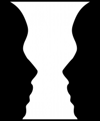
Dividing visual displays into figure (thing being looked at) and ground (background against which it stands); fundamental way in which people organize visual perceptions
|
|
|
Similarity |
People tend to group stimuli that are similar |
|
|
Proximity |
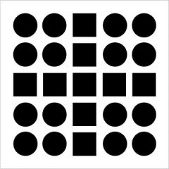
Elements that are close to one another tend to be grouped together |
|
|
Closure |
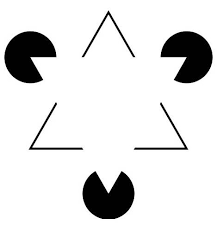
Viewers tend to supply missing elements to close or complete a familiar figure |
|
|
Continuity |

Viewers tend to see elements in ways that produce smooth continuation |
|
|
Perceptual Consistancies |
Recognize stimuli under varying conditions (shape, brightness, size) |
|
|
Depth/Distance Perception |
Image on the retina is 2 dimensional but we live in and perceive a 3-D world; possible through binocular and monocular cues |
|
|
Binocular Cues |
Cues from both eyes together; Retinal disparity (objects project images to slightly different location on the right and left retinas, so the right and left eyes see slightly different views of the object); Convergence (muscles move closer/farther to adapt to depth and distance) |
|
|
Monocular Cues |
Cues from a single eye; Motion parallax (images of objects at different distances moving across the retina at different rates); Accomodation; Pictorial depth cues (clues about a distance that can be given in a flat picture) |
|
|
Aspects of Pictorial Depth Cues |
Light and shadow, linear perspective, interposition, height in horizontal plane, texture, clarity, relative size |