![]()
![]()
![]()
Use LEFT and RIGHT arrow keys to navigate between flashcards;
Use UP and DOWN arrow keys to flip the card;
H to show hint;
A reads text to speech;
38 Cards in this Set
- Front
- Back
|
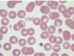
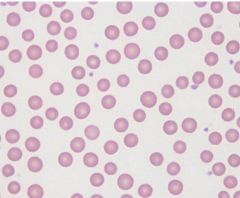
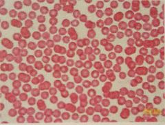
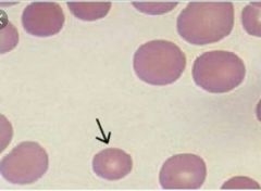
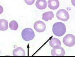
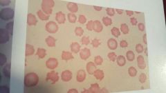
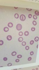
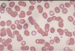
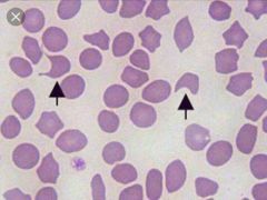
Canine Erythrocyte , also Normocytic Erythrocyte Biconcave disk Largest of the RBC'S in domestic species Uniform in size Central pallor appears pale |
|
|
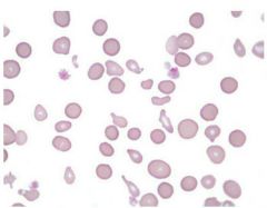
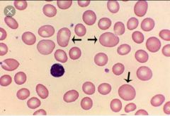
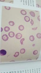
Feline Erythrocyte Large RBC but smaller than the dog Mild anisicytosis (variation in cell size) Limited central pallor Crenation is common Howells Jolly bodies (nuclear remnants) may occur up to 1% of RBC'S Rouleaux formation may be noted |
|
|
Equine Erythrocyte Nearly flat, non-nucleated, homogeneous Similar in size to feline RBC Lack central pallor Rouleaux formation is common |
|
|
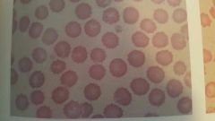
Bovine (Cow) Erythrocyte Round non-nucleated, homogeneous A little smaller than the horse RBC Common anisocytosis Limited central pallor Crenation is common |
|
|
Ovine Sheep Erythrocytes Small, round, non-nucleated Similar to cows RBCs but smaller Common anisocytosis Common poikilocytosis (variation in shape) Limited central pallor |
|
|
Caprine Goat ErythrocytesSmallest RBCs of domestic speciesCommon anisocytosisCommon poikilocytosisMinimal central pallor |
|
|
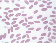
Camelidae (camel, Llama, alpaca etc.) Erythrocytes Oval or elliptical in shape and thin Non- nucleotide and homogeneous No central pallor |
|
|
Birds/Reptiles/Amphibians/Fish Erythrocytes Oval/Elongated Contain a central oval nucleus Hint: Look like eggs and they all lay eggs. |
|
|
Microcytes Smaller than normal |
|
|
Macrocytes Larger than normal |
|
|
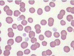
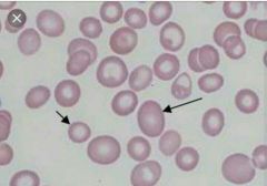
Anisocytosis Variation in cell size |
|
|
Hypochromic/Hypochromasia Increased central pallor Decreased cytoplasmic stain intensity Caused by decreased/insufficient hemoglobin in RBC Common cause- iron deficiency or lead toxicities |
|
|
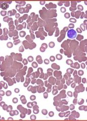
Polychromic/Polychromasia Blue-gray RBC with residual RNA Usually macrocytic Romanowsky stain- polychromatophils (aka reticulocytes) NMB stain- Reticulocytes (aka polychromatophils) A few are normal in dogs and cats Less common in cows Rarely seen in horses Increased numbers indicate a regenerative response to anemia. |
|
|
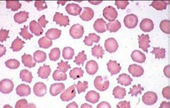
Poikilocytosis General term for abnormally shaped cells Indicates cell degradation/ defects or toxic damage |
|
|
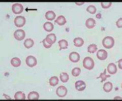
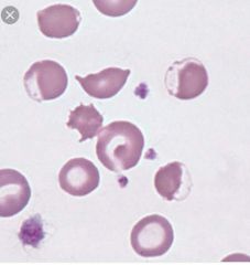
Echinocytes Burr looking Type 1 cause- Crenation Type 2 causes (true echinocytes) - renal disease and Rattlesnake bites |
|
|
Burr cells Elongated echinocytes |
|
|
Acanthocytes Look like a blood splatter Formed from alterations in cholesterol and phospholipids Seen in: Kidney or liver disease, hemangiosarcoma, lymphoma, cancer |
|
|
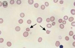
Spherocytes Microcytic and lack central pallor Densely stained Hemoglobin Seen in: immune mediated hemolytic anemia (IMHA) and post blood transfusions |
|
|
Eccentrocytes Oxidative injury Hemoglobin concentrated at one end and the opposite end is unstained Shows injury to RBC membrane Toxic injestion: onions, propylene glycol, and acetaminophen |
|
|
Target cells Dark center and outer area Usually an artifact/nonspecific change If seen in large numbers could be Regenerative Anemia and Liver Disease |
|
|
Torocytes Punched out cells Usually artifact of slide preparation |
|
|
Schisocytes Mechanical Fragmentation RBC fragments due to cell membrane damage Seen in: trauma cases, Disseminated Intravascular Coagulation (DIC) [run into microclots. Sort of like a rocky river ripping off pieces. |
|
|
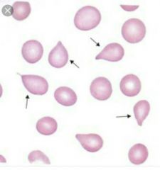
Dacryocytes Mechanical Fragmentation Tear drop shaped Artifact of slide preparation |
|
|
Reticulocytes Regeneration Last stage of erythropoiesis Cats have 2 forms aggregate and punctate Aggregate=reticulum in clumps Punctate= only have a few isolated dots of reticulum |
|
|
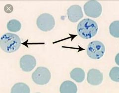
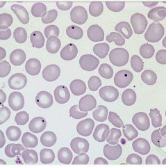
Basophilic Stippling Regenerative Use Romanowsky stains Abnormal clumping of RNA appearing as small blue dots Seen in lead poisoning Regenerative response is more common in ruminants |
|
|
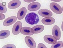
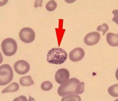
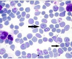
Nucleated RBC Regenerative Metarubicyte cell stage before reticulocyte Look like wbcs |
|
|
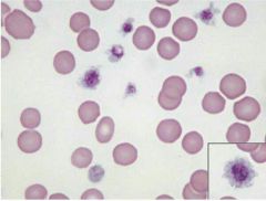
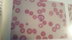
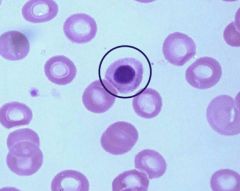
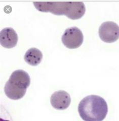
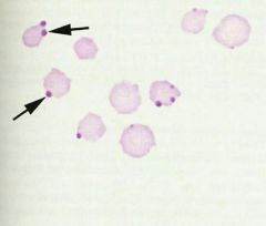
Howell-Jolly Bodies Small spherical nuclear fragment in RBC Shows accelerated RBC production/regeneration Found in splenectomy patients |
|
|
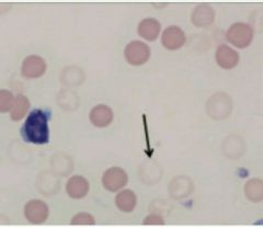
Ghost cells Immune Mediated Damage Very pale and small Caused by Intravascular hemolysis If it is an artifact it will appear as a red smudge |
|
|
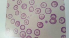
Rouleaux Formation Immune Mediated Damage Stacked coins look Common in horses Occasionally in cats Increased fibrinogen make RBCs sticky Seen in inflammatory diseases |
|
|
Agglutination Immune Mediated Damage Clumping of RBCs Antigens on RBCs interact with antibodies causing agglutination Macroscopic or micro Seen in IMHA Not normal never normal! |
|
|
Heinz bodies Oxidative injury to hgb Nose like projection Common in up at 10% of cats RBC Associated with hemolytic anemia in all species Caused by Zinc, Acetaminophen, Onion, maple leaves toxicity in horses, propophol in cats |
|
|
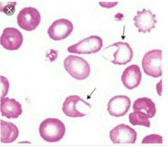
Blister Cell Metabolic/Membrane Disorders Intact blister like membrane structures Precursor cell to the keratocyte Causes: artifact, vascular damage, excess coagulation, anemia |
|
|
Keratocyte Metabolic/ Membrane Disorder Helmet or horns Causes: artifact, vascular damage, excess coagulation, anemia Blister cell is formed in RBC membrane and it ruptures |
|
|
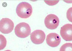
Babesia Canis RBC parasite Tear dropped or pear shaped intracellular organism Transmitted by brown dog tick Clinical signs: severe anemia, fever, icterus, hemolysis, hemoglobinuria, shock |
|
|
Babesia gibsoni RBC parasite Small ring shaped intracellular parasite Transmitted to dogs by ticks, infected blood transfusions, and dog bites Greyhounds and Pit Bulls most susceptible, may be asymptomatic carriers Difficult to diagnose, genetic testing |
|
|
Mycoplasma haemofelis Haemobartonella RBC parasite Epicellular parasite (occurs within depressions on cell surface) Feline infectious anemia Blue staining cocci, rings, or rods Transmitted by fleas and or ticks Clinical signs: mild to severe anemia, fever and icterus |
|
|
Mycoplasma canis Formerly known as H. canis RBC parasite Very small Epicellular parasite (occurs within depressions on the cell surface) Coccoid or rod shaped Found individually or in groups on RBCs Often forms chains across the RBCs |
|
|
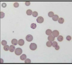
Anaplasma marginale RBC parasite Transmitted by ticks to cattle Clinical signs: anemia, icterus, depression, fever, abortions Round oval basophilic inclusions Located at periphery of cell |