![]()
![]()
![]()
Use LEFT and RIGHT arrow keys to navigate between flashcards;
Use UP and DOWN arrow keys to flip the card;
H to show hint;
A reads text to speech;
83 Cards in this Set
- Front
- Back
|
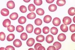
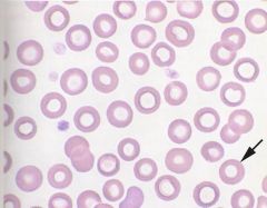
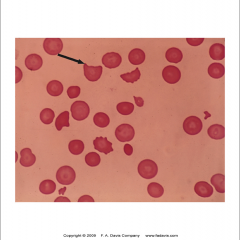
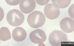
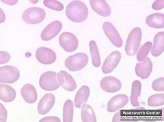
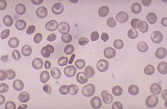
Normochromic, normocytic erythrocytes (normal) |
|
|
Normochromic, normocytic erythrocytes (normal) |
|
|
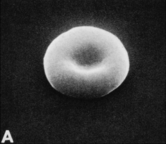
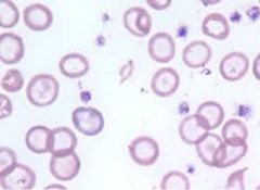
Normochromic, normocytic erythrocytes |
|
|
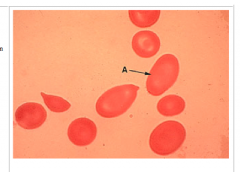
A.Normochromic, normocytic erythrocytes |
|
|
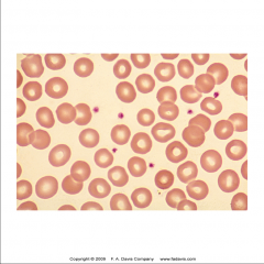
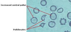
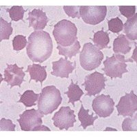
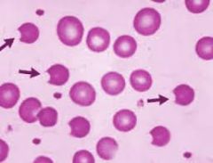
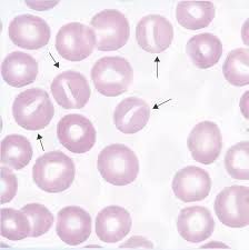
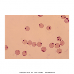
Hypochromia is characterized by an increase in central pallor. It is said to be present when greater than 1/2 of the cell diameter has central pallor. Hypochromia is caused by a deficiency in the production of hemoglobin, which is usually the result of iron deficiency caused by chronic hemorrhage.
|
|
|
hypochromasia |
|
|
hypochromasia |
|
|
hypochromasia |
|
|
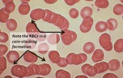
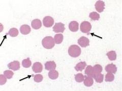
whenthe red cells are stacked in piles as they are here, this formation is called “rouleaux”. Itis caused by high protein concentrations in plasma. |
|
|
rouleaux |
|
|
rouleaux |
|
|
Agglutination occurs if an antigen is mixed with its corresponding antibody called isoagglutinin. This term is commonly used in blood grouping. |
|
|
Agglutination |
|
|
Agglutination |
|
|
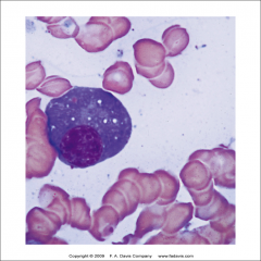
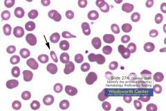
metarubricytes,with their dark purple, pyknoticnuclei Whenmetarubricytes areseen in PB, it is an indication that the BM is releasing RBCs before they arefully mature. This can be seen in cases of severe anemia, no matter what thecause is. |
|
|
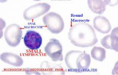
amegoloblastic metarubricyte is also observed. It kind of looks like a small lymphocyte but not really! Normally the nucleus of the metarubricyte would be smaller but this cell has delayed nuclear maturation so it is larger |
|
|
metarubricyte |
|
|
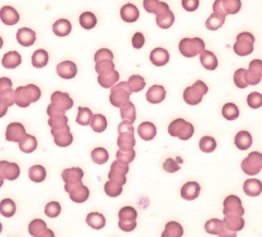
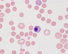
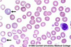
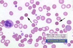
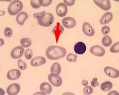
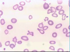
polychromatophilic erythrocytes which are larger than normal RBCs
|
|
|
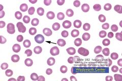
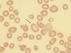
In the lower right quadrant of this photo is a polychromatophilic erythrocyte, among other, normal mature red blood cells, indicated by the white arrow. Please remember, this cell can only be called a reticulocyte when it is stained with a special stain. Note that this cell is larger and has a bluish tint as compared to the surrounding mature erythrocytes
|
|
|
polychromatophilic |
|

|
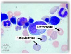
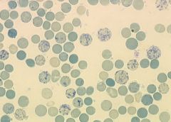
The cells can be stained by a special stain known as a reticulocyte stain – using this stain, the cells are called reticulocytes. When it is formed, the reticulocyte contains only about 2/3 the amount of hemoglobin it will contain as a mature erythrocyte. Reticulocytes remain in the bone marrow 2 to 3 days after formation and circulate in the blood 1 to 2 days before maturing into an erythrocyte.
|
|
|
reticulocyte |
|
|
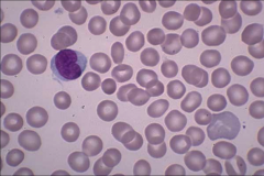
reticulocyte (top left) |
|
|
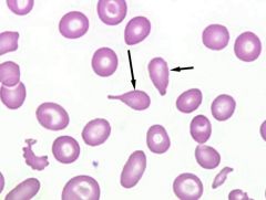
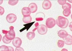
Spherocytes are cells with a reduced surface to volume ratio. The survival time of these redblood cells in the bloodstream is greatly shortened because they have lost their biconcave shape and they can’t squeeze through small areas very well.
|
|
|
Spherocytes They can be the result of genetic abnormalities but they can also result when a macrophage in the spleen phagocytoses part of the membrane of a passing red cell that isn’t flexible enough to slip by without notice! |
|
|
Spherocyte |
|
|
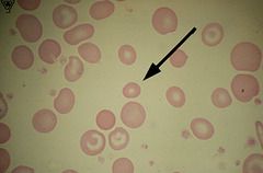
Microcyte unusually small red blood cell, associated with certain anemias. still have biconcave |
|
|
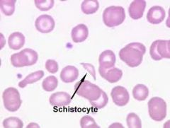
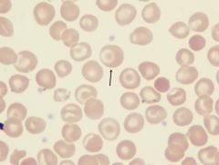
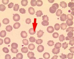
Schistocytes are typically irregularly shaped, jagged, and have two pointed ends
|
|
|
A true schistocyte does not have central pallor.
|
|
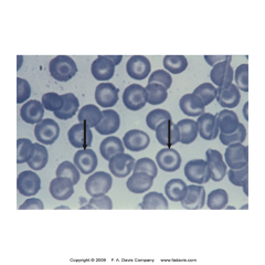
|
target cell |
|
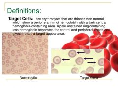
|
target cell |
|
|
target cell |
|
|
target cell |
|
|
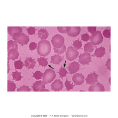
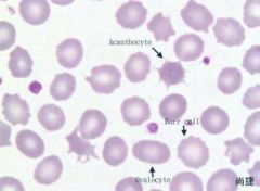
acanthocyte spiked cell membrane, due to abnormal thorny projections
|
|
|
acanthocyte |
|
|
Burr cell (echinocyte) Echinocytes, more commonly referred to as burr cells, are reversible, meaning that this alteration can be the result of the cell's environment, pH of the medium (including the glass slides on which blood smears are made), the metabolic state of the cell and the use of some chemical substances. |
|
|
burr cell |
|
|
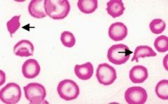
Bitecells are red blood cells that result when the removal of a part of the cellmembrane leads to a permanent indentation in the remaining cell. Again, thesurvival time of these cells is greatly shortened because they have lost theirbiconcave shape and they are no longer flexible enough to survive
|
|
|
bite |
|
|
bite |
|
|
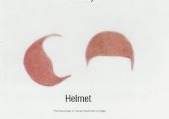
helmet |
|
|
helmet |
|
|
themacroovalocyte --it looks smaller in this picture but as you already know, macrocytes arelarger than normal erythrocytes
|
|
|
macrocyte |
|
|
Macro ovalocytes are enlarged, oval-shaped erythrocytes (red blood cells). They are not seen in healthy blood, and are most commonly seen in megaloblastic anemia
|
|
|
macroovalocyte |
|
|
Stomatocytes are red blood cells with an oval or rectangular area of central pallor
|
|
|
stomatocyte |
|

|
stomatocyte |
|
|
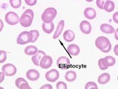
Elliptocytes are red blood cells that are oval or cigar shaped. They may be found in various anemias, but are found in large amounts in hereditary elliptocytosis
|
|
|
elliptocyte |
|
|
elliptocyte |
|
|
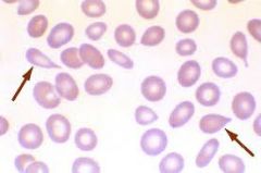
A dacrocyte (or dacryocyte) is a type of poikilocyte that is shaped like a teardrop ( a "teardrop cell")
|
|
|
teardrop cell |
|
|
teardrop cell |
|
|
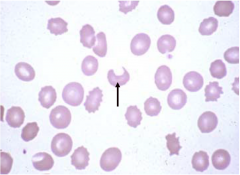
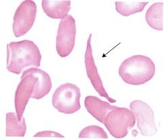
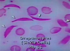
A crescent-shaped, holly-leaf-/scythe-like RBC caused by polymerisation or sickling of haemoglobin (Hb S) or other “sickling” Hbs, which occurs more readily with decreased pH, decreased O2, increased temperature and increased osmolarity.
|
|
|
Drepanocyte |
|
|
Depranocyte |
|
|
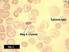
in the center of the field is a rectangular RBC that is indicative of a hemoglobin C crystal, which is also characteristic for hemoglobin C disease.
|
|
|
hemoglobin c crystals |
|
|
Hemoglobin c crystals |
|
|
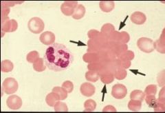
SC crystals (single arrow
|
|
|
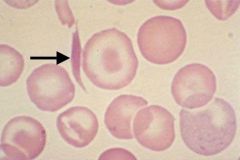
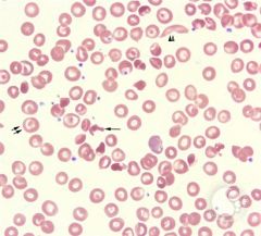
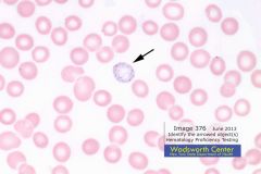
On the right side, the arrow points to a cell with a Howell-Jolly body. Remember,these are fragments of the nucleus that have been left behind in the red blood cell when it exits the bone marrow. They stain dark purple with the Wright stain because they contain DNA
|
|
|
howell jolly body |
|
|
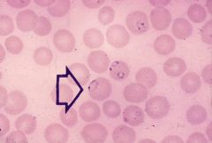
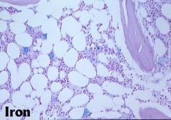
Iron can be seen in red blood cells in a smear when it is stained with Prussian blue, as seen here (iron granules are called Pappenheimerbodies when observed in a Wright stain
|
|
|
pappenheimer bodies |
|
|
pappenheimer bodies |
|
|
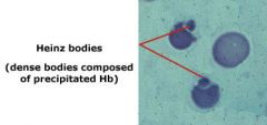
These are Heinz bodies. They are abnormal inclusion bodies found in red cells and they are the result of denatured or precipitated hemoglobin. Heinz bodies cannot be observed in a normal peripheral blood smear stained with Wright stain.They are only seen with special stains such as crystal violet or brilliant cresyl blue.
|
|
|
heinz bodies |
|
|
heinz bodies |
|
|
heinz bodies |
|
|
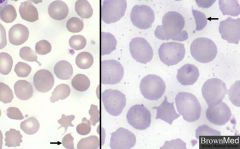
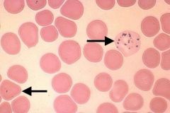
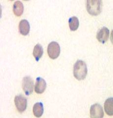
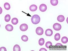
basophilic stippling is due to the presence of ribosomes inside the red blood cell. It is considered a characteristic feature of lead poisoning, but is seen in other diseases such as alcoholism, thalassemias, and megaloblastic anemias.
|
|
|
basophilic stippling |
|
|
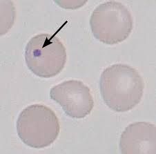
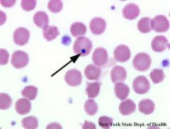
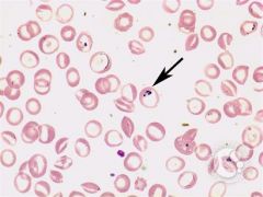
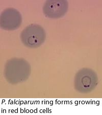
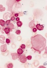
malarial forms |
|
|
malarial forms |
|
|
malarial forms |
|
|
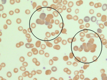
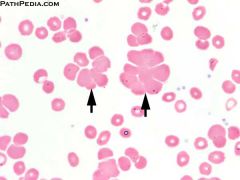
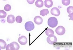
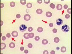
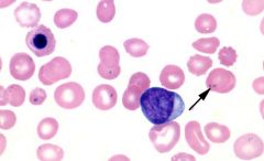
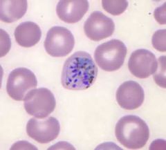
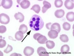
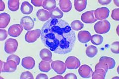
hypersegmentation of neutrophils |
|
|
hypersegmentation of neutrophils |
|
|
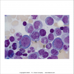
iron stain bone marrow |
|

|
iron stain bone marrow |
|
|
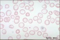
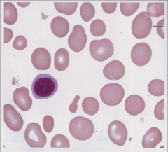
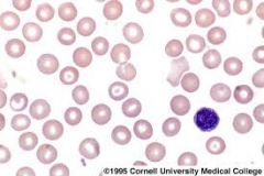
peripheral blood |
|
|
peripheral blood |
|
|
reticulocyte stain methylene blue |