Reading...
![]()
Play button
![]()
Play button
![]()
Use LEFT and RIGHT arrow keys to navigate between flashcards;
Use UP and DOWN arrow keys to flip the card;
H to show hint;
A reads text to speech;
14 Cards in this Set
- Front
- Back
|
Example of barium Follow through
|

|
|
|
Example of small bowel enema
|

|
|
|
What is malabsorption?
|
Abnormal absorption of fat, water, protein, carbohydrates from small bowel
|
|
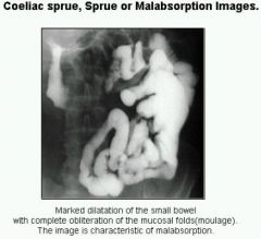
Radiographic features of malabsorption:
|
•Dilatation of bowl loops
•Diluted barium (mixes with watery bowel content) •Flocculated barium : barium aggregates into particles (mainly seen with older barium suspensions, rarely occurs with new agents) •Slow transit •Segmentation of barium (lack of continuous column) •Moulage pattern: featureless barium collection •Hidebound pattern: valvulae thinner, closer together, wrinkled looking |
|
|
What is Coeliac Sprue?
|
Its is a disease hat affects the mucosa of the small bowel and the submucosa, muscularis and serosa are usually intact.
it is hereditary with genetic transmission of an inborn error or metabolism |
|
|
What are common symptoms of coeliac sprue?
|
diarrhoea, flatulence, weight loss and weakness
|
|
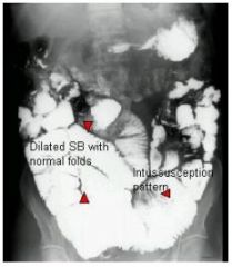
Radiographic features of coeliac sprue:
|
•dilatation of small bowel is the most typical finding
•Segmentation •Moulage sign: rare •Transient intussusceptions (coiled spring) are typical •Decreased fold pattern of jejunum •Prominent ileal fold pattern (ileum appears like jejunum) •Increased secretions: flocculation with older barium suspensions •Increased incidence of malignancy, aggressive lymphoma, carcinoma |
|
|
Characteristic image of malabsorption
|
|
|
|
Fissure ulcer - a characteristic of Chrohn's disease
|

|
|
|
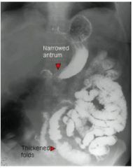
Radiographic features: types of lesions
|
– Thickening of folds
– Nodular pattern – String sign: tubular narrowing of intestinal lumen |
|
|
Radiographic features: ulceration
|
– Aphthoid ulcers are early lesions & correspond to mucosal erosions
– Ulcers are irregularly scattered through out GI tract & are interspersed with normal mucosa – Ulcerations grow and fuse with each other in linear fashion and with intervening oedematous mucosa |
|
|
Radiographic features: Crohn’s disease at advanced stages
|
Fatty thickening and/or retraction of the mesentery, mass effect may separate bowel loops or make loops display a concentric shape
|
|
|
Radiographic features: Fibrosis and scarring may result in
|
– Pseudodiverticula
– Rigid, featureless bowel – Stricutres and obstruction – Foreshortening of bowel |
|
|
Complications of Chrohn's Disease (RIFTA)
|
-Recurrence (after surgery)
-incresed incidence of malignancies (lymphoma, GIT tumours) -toxic megacolon -fistulas abscess formation |