Reading...
![]()
Play button
![]()
Play button
![]()
Use LEFT and RIGHT arrow keys to navigate between flashcards;
Use UP and DOWN arrow keys to flip the card;
H to show hint;
A reads text to speech;
68 Cards in this Set
- Front
- Back
|
Show the following landmarks for the knee and lower leg region and give the soft tissues (muscle, tendon, ligaments) attached:
|
a) Tibial tuberosity
b) Inferior pole of patella c) Superior pole of patella d) Patella tendon e) Quadriceps tendon f) Fibula head g) Tibiofibular joint h) Common fibular (peroneal) nerve i) Popliteal artery j) Lateral collateral ligament k) Medial collateral ligament l) Pes anserinus bursa and tendons m) Site of bursa complex around knee n) Femoral condyles o) Tibial plateaus and joint space - anterior, medial and lateral p) Adductor tubercle q) Semimembranosis tendon and muscle r) Semitendinosus tendon and muscle s) Sartorius tendon and muscle t) Gracilis tendon and muscle u) Biceps femoris tendon and muscle v) Gastrocnemius tendons w) Vastus medialis muscle x) Vastus lateralis muscle y) Rectus femoris muscle z) Vastus intermedius muscle aa) Iliotibial band insertions |
|
|
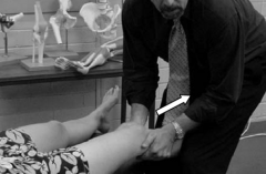
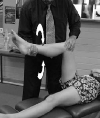
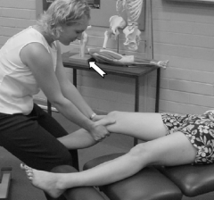
Tibiofemoral joint
long axis distraction |
|
|
|
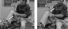
A-P/P-A glide (90o flexion)
|
|
|
|
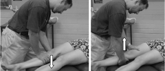
A-P/P-A glide extension
|
|
|
|
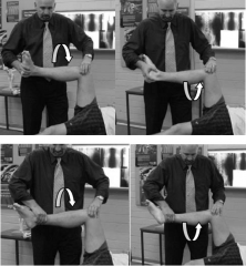
Internal/external rotation (90o flexion)
|
|
|
|
Internal/external rotation (extension)
|
|
|
|
M-L/L-M glide (extension)
|
|
|
|
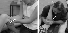
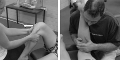
Tibiofibular: (Proximal)
A-P/P-A glide (90o flexion) |
|
|
|
A-P/P-A glide (extension)
|
|
|
|
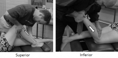
S-I/I-S glide (90o flexion)
|
|
|
|
S-I/I-S glide (extension)
|
|
|
|
Patellofemoral:
I-S/S-I glide |
|
|
|
M-L/L-M glide
|
|
|
|
clockwise/counterclockwise rotation
|
|
|
|
Rectus femoris
|
|
|
|
Quadriceps group
|
|
|
|
Adductors
|
|
|
|
Sartorius
|
|
|
|
Tensor fascia lata
|
|
|
|
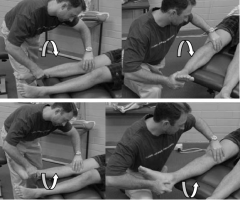
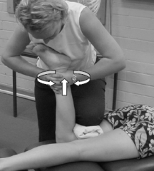
Popliteus
|
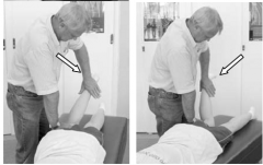
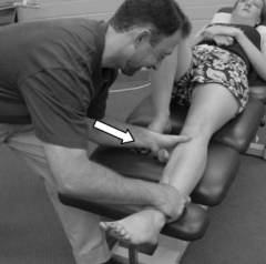
T.P.: Prone, knee flexed 90º, tibia medially rotated on femur.
TEST: Attempt to rotate tibia laterally on femur. OBS: Knee flexion or extension, rotation of femur at hip. |
|
|
Hamstring group
|
|
|
|
Lateral hamstring
Medial hamstring |
|
|
|
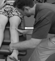
Gracilis
|
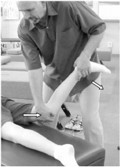
Pressure is directed against the posterior medial aspect of the distal leg in a direction of knee extension and slightly lateral. Thigh must be kept in position of extension and abduction.
|
|
|
General Ortho tests
Bounce Home test |
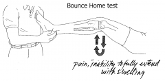
Flex knee then let go
Provocative Test for ACL, PCL and Meniscus |
|
|
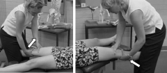
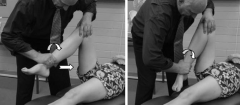
Collateral ligaments
valgus stress tests |
In neutral (extension) or with 30 degrees of flexion
- L-M force (VALGUS) applied over knee joint Place your hand either on the femur or the tibia when applying valgus force to be more specific |
|
|
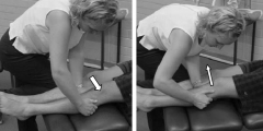
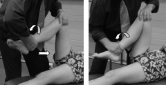
Varus Stress test
|
To test the lateral collateral ligament apply VARUS stress to the knee joint on the lateral side
|
|
|
Cruciate ligaments:
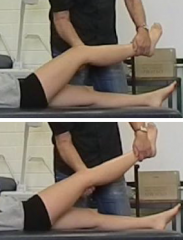
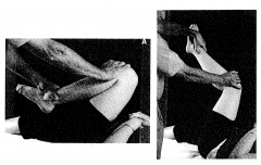
anterior drawer sign |
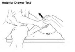
Anterior cruciate ligament
Positive: Instability, increased mobility |
|
|
posterior drawer sign
|
Posterior cruciate ligament
Positive: Instability, increased mobility |
|
|
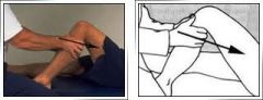
Lachman’s test
|
Anterior cruciate (Better test- Gold standard)
Patient knee flexed to 30 degree, so as to be in the open-packed position Support the patient femur and apply a P-A force to the back of the knee |
|
|
Reverse lachman’s
|
Posterior cruciate ligament
- Put the patient knee in 30 degree flexion, open packed position - Support at the femur and on the tibia and apply a force in the A-P direction |
|
|
Pivot shift test
|
IR Tibia on fully extended knee with VALGUS stress
- Clunks about 20o knee flexion - Deficiency of ACL (grade 1 or 2 tear) |
|
|
Reverse pivot shift test
|
ER on fully flexed knee with VALGUS stress
Extent- Clunk at about 40o flexion - Deficiency of PCL (grade 1 or 2 tear) |
|
|
Slocum’s test
|
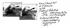
Knee flexed to 90o P-A Drawer with:
- Internal rotation- ACL, Lateral capsule/Lig, ITB - External rotation- ACL, Medial capsule/Lig |
|
|
Posterior sag
|
PCL tear
Look for dropping of leg |
|
|
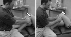
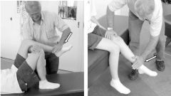
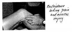
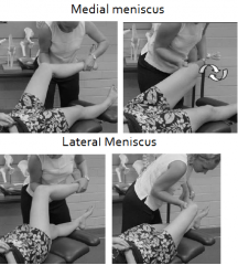
Menisci:
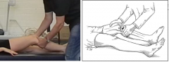
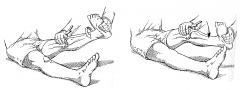
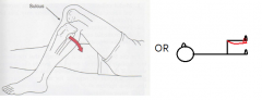
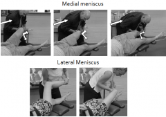
McMurray’s test- Loose fragment |
Medial Meniscus: (PIC) - Hip flexed at 90 degrees and tibia flexed at knee with external rotation,
Lateral Meniscus: - Hip flexed at 90 degrees and tibia flexed at knee with internal rotation, - Reproduces a pain or click in joint space |
|
|
Joint line tenderness
|
|
|
|
Steinmann’s tenderness displacement test
|
With knee in FLEXION palpate most tender point on joint line. Pain disappears if meniscus lesion as meniscus move forward when you extend the knee.
Medial pain is elicited on lateral rotation, and lateral pain is elicited on medial rotation. |
|
|
Apley’s test grind/distraction
|
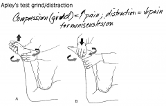
- Compression (grind)= increased pain
- Distraction= decreased pain - Indication of meniscus lesion |
|
|
Thessaly test
|
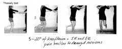
Produce pain on side where there is pain on damaged meniscus side
|
|
|
Effusion:
Patellar tap test/ Bulge or sweep (Milking) |
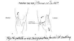
- Tap on top of patella, look for radiating pain
- Fluid movement from milking/ sweeping - Ppl with large adipose tissue build up and damage to knee region |
|
|
Patella ballottement
|
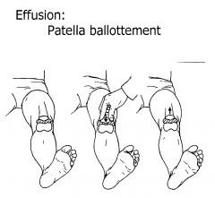
Push patella into joint and release suddenly patella seen to rise visibly. Positive test if patella slowly floats
|
|
|
Patella:
Apprehension test |
Pain, uncomfortable (Push M-L) The knee flexed to 30°
If the patient feels the patella is going to dislocate, the patient will contract the quadriceps muscles to bring the patella back "into line." |
|
|
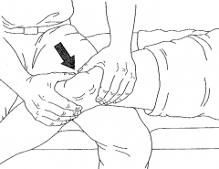
Grinding test/Fouchet’s
|
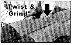
Twist and grind patella with compression- Positive if pain present
|
|
|
Clarks
|
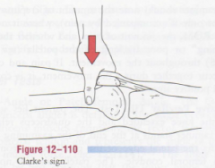
Pain on compression and Quads contraction
|
|
|
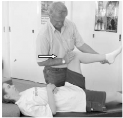
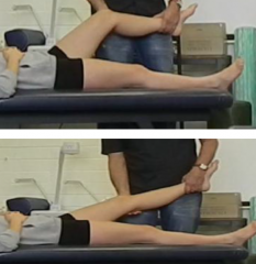
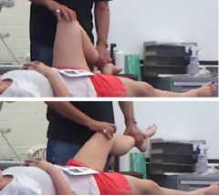
5 Perform the following functional tests for the knee and lower leg region:
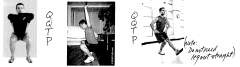
a) Screw Home Mechanism + VMO assessment (Timing, Quality and Quantity, Pain) |
Start with patient in sitting position. (Knees flexed).
- Place a pen mark on the tibial tuberosity. - Place a pen mark on the inferior pole of the patella. - The patient is asked to actively extend the knee. - Observes for (Lateral movement/External rotation of the tibia) with Slight medial/internal rotation of the patella (pulled by the Vastus Medius) Palpate- Patella, Tibial tiberosity, Vastus medialis Factors- DJD, Tracking syndrome, knee reco or replacement, condromelasia Throughout your observation and palapation you are assessing QQTP as a whole and comparing side to side. Look for asymmetry. The most reliable indicator is the Vastus Medius contraction Screw Home Mechanism: Tiba rotates externally, Vastus Medialis which counteracts the lateral pull of Vastus lateralis, ITB and Rectus Femoris. VM has a controlling action |
|
|
b) Squat assessment – Heels on/off (TQQP)
|
|
|
|
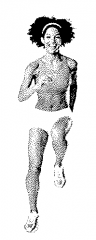
c) One legged hop
|
|
|
|
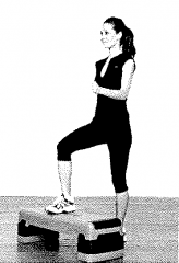
d) Step up-step down
|
|
|
|
e) Functional genu valgus/varus assessment
|
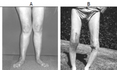
A- Valgus
B- Varus |
|
|
f) Assess the Q angle
|
1. Patient standing in neutral position.
2. Mark the middle of the patella. 3. Draw a line from the mid-tibial tuberosity to the patella. 4. Draw a line from the ASIS to the patella. 5. Measure the inside angle between the two lines below the patella intersection. o Males will have 5-15 degrees o Females will have a larger Q angle at 10-20 degrees |
|
|
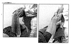
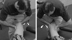
McMurray’s Modification Technique #1Tibial knee external rotation and abduction (slow manoeuvre with traction)
|
Displacement/ Minor tears of medial meniscus.
The foot and leg are externally rotated. VALGUS force, traction applied Lateral meniscus- Contralateral, tibial internal rotation and adduction force (varus) combined with the traction. |
|
|
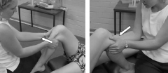
McMurray’s Modification Technique #2 Tibial knee external rotation and abduction (circumduction with thrust)
|
Displacement/ Minor tears of medial meniscus.
The foot and leg are externally rotated. VALGUS force, Thrust applied Lateral meniscus- Contralateral, tibial internal rotation and adduction force (varus) combined with the thrust. |
|
|
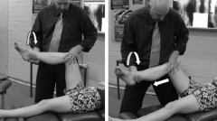
Alternating circumduction and extension
|
Traction Applied (side-to-side movement at the end)
Medial meniscus- the knee in valgus position and then circumducted medially with extension Lateral meniscus- The knee is placed is a varus position and circumducted laterally. with extension |
|
|
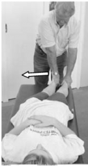
A-P drawer technique
|
Can be applied as set thrust 6-12 times
OR A-P movement 30-60 times |
|
|
Tibiofemoral circumduction technique
|
|
|
|
Tibial rotation technique
|
|
|
|
Apley’s modification Technique
|
|
|
|
Gertler’s technique
|
Internal and external rotation with traction
|
|
|
Popliteal (flexion) stretch (prone/supine)
|
|
|
|
Medial tibia subluxation adjustment technique
|
|
|
|
Lateral tibia subluxation adjustment technique
|
|
|
|
External rotation subluxation tibia adjustment [external rotary knee adjustment]
|
Run up technique- thrust IR at end of motion
|
|
|
Internal rotation subluxation tibia adjustment [internal rotary knee adjustment)
|
Run up technique- thrust ER at end of motion
|
|
|
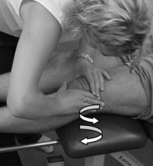
Tibial lift technique
|
Loss of P-A glide or distraction (locked knee)
|
|
|
Posterior fibula subluxation technique (supine and prone)
|
|
|
|
Posterior tibia subluxation technique (supine and prone)
|
|
|
|
Superior/inferior fibula subluxation technique
|
|
|
|
Patella adjustment technique
|
|