Reading...
![]()
Play button
![]()
Play button
![]()
Use LEFT and RIGHT arrow keys to navigate between flashcards;
Use UP and DOWN arrow keys to flip the card;
H to show hint;
A reads text to speech;
13 Cards in this Set
- Front
- Back
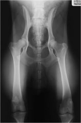
Bones: Ilium, ischium, pubis, acetabular bone, sacrum
Joints: Sacroiliac joint, coxofemoral joint Landmarks: Obturator formane, iliopubic eminence, ischiatic tuberosity, ischiatic arches |
|
|

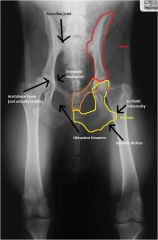
Bones: Ilium, ischium, pubis, sacrum, acetabular bone
Joints: Coxofemoral joint Landmarks: Ischiatic tuberosity, obturator foramen |

|
|
Bones: Ilium, ischium, pubis, sacrum, acetabular bone
Joints: Coxofemoral joint Landmarks: Ischiatic tuberosity, obturator foramen |
|
|
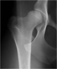
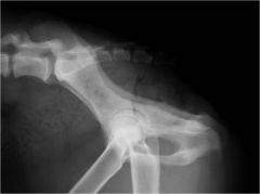
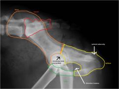
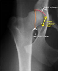
Bones: Pelvis, Femur
Joints: Coxofemoral joint Landmarks: Cranial acetabular rim, dorsal acetabular rim, ventral acetabular notch |
|
|
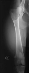
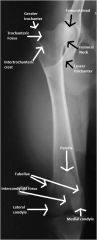
Bones: Distal Pelvis, Femur, Proximal Tibia
Joints: Coxofemoral joint, Femorotibial joint Landmarks: Femoral head, femoral neck, greater and lesser trochanters, trochanteric fossa, intertrochanteric crest, patella, fabellae, intercondyloid fossa, medial and lateral condyles |
|
|
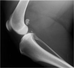
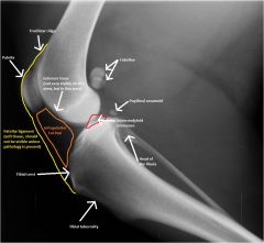
Bones: Distal femur, proximal tibia and fibula
Joint: Femorotibial joint Landmarks: Patella, fabellae, popliteal sesamoid, trochlear ridge, extensor fossa, infrapatellar fat pad, patellar ligament, intercondyloid eminence, head of the fibula, tibial crest, tibial tuberosity |
|
|
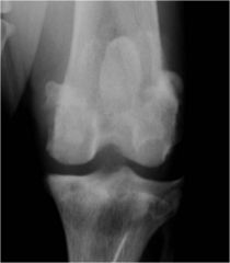
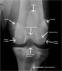
Bones: Distal femur, proximal tibia and fibula
Joint: Femorotibial joint Landmarks: Patella, fabellae, extensor fossa, intercondyloid fossa, medial and lateral condyle, intercondylar eminence, tibial tuberosity |
|
|
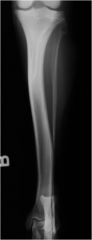
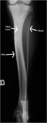
Bones: Distal femur, Tibia, Fibula, tarsal bones
Joints: Femorotibial, tibiotarsal Landmarks: Tibial crest |
|
|
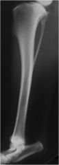
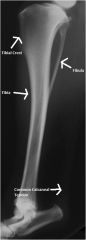
Bones: Distal femur, Tibia, Fibula, tarsal bones
Joints: Femorotibial, tibiotarsal Landmarks: Tibial crest, Common calcaneal tendon |
|
|

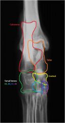
Bones: Talus, Calcaneus, Central, Tarsal bones 1-4
Joints: Tibiotarsal, proximal and distal intertarsal, tarsometatarsal |
|
|
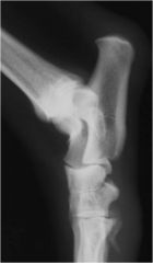
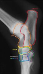
Bones: Talus, Calcaneous, Central, Tarsal bones 1-4
Joints: Tibiotarsal, proximal and distal intertarsal, tarsometatarsal |
|
|
|
What features distinguish medial from lateral on the small animal hind limb?
|
1. Orientation to the pelvis.
2. Greater trochanter is lateral, lesser trochanter is medial. 3. Extensor fossa is on the lateral femoral condyle 4. Fibula is on the lateral side. 5. 4th tarsal bone is largest and most lateral. |
|
|
The extensor fossa of the femur can be mistaken for what kind of lesion?
|
Osteochondrosis lesion
|