![]()
![]()
![]()
Use LEFT and RIGHT arrow keys to navigate between flashcards;
Use UP and DOWN arrow keys to flip the card;
H to show hint;
A reads text to speech;
29 Cards in this Set
- Front
- Back
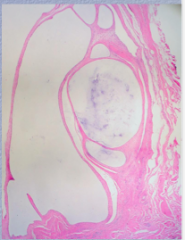
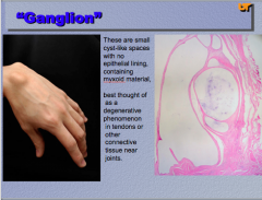
What is the term for these spaces? Do they contain an epithelial lining? Mixoid material?
Degenerative phenomenon in _______ or other connective tissue near _________. |
|
|
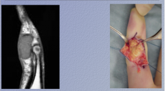
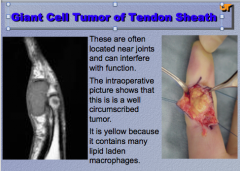
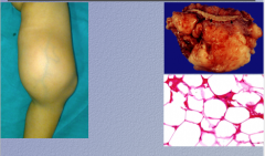
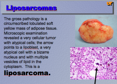
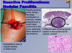
What is shown here? Where are they often located? Why is the tumor yellow? |
|
|
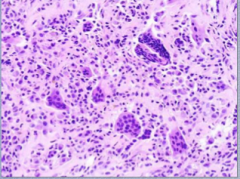
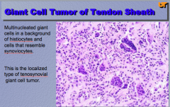
What cells do you see? Background of what that resembles what?
What is this a localized type of? |
|
|
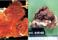
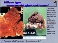
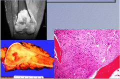
What type of tumor is shown? Where is it usually located? What causes the red color? What happens to the joint? |
|
|
FAT FAT FAT! What is this? |
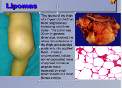
Lipoma |
|
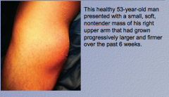
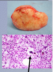
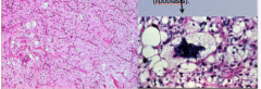
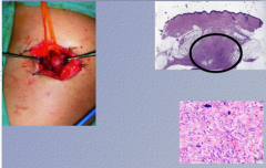
What is this? |
Liposarcoma |
|
What is the condition? |
|
|
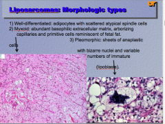
What are the three morphologic types of liposarcoma? Which two types are shown here? |
|
|
What is the disease? Where does it usually start? How does it normally present? Where? |
|
|
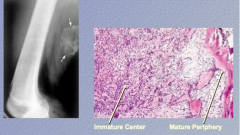
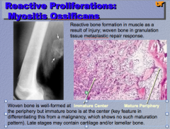
Woven bone at periphery. Immature bone at center. What is the diagnosis? Is it malignant? How do you know? |
|
|
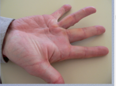
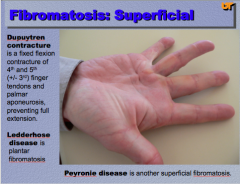
What is the term for a fixed flexion contracture of 4th and 5th finger tendons and palmar aponeurosis? What is plantar fibromatosis? What is superficial fibromatosis?
|
|
|
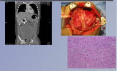
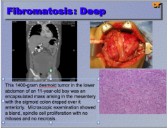
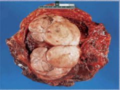
What type of tumor? Spindle cell proliferation with no mitoses and no necrosis. |
|
|
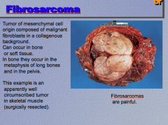
Painful, mesenchymal cell origin, fibroblasts in collagenous background. Bone or soft tissue. Where do they occur in bone? What is the diagnosis? |
|
|
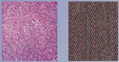
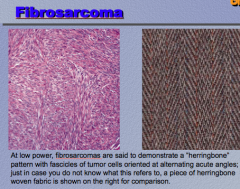
What is the pattern? |
|
|
|
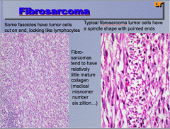
Fibrosarcomas tend to have relatively little mature what...?
What do some fascicles look like? What is the shape of a typical fibrosarcoma tumor cell? |
|
|
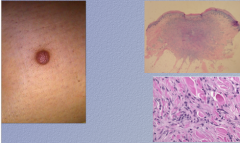
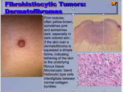
Firm, yellow brown, dimple formed if skin squeezed. Histology = histolytic type cells between normal collagen bundles Diagnosis? |
|
|
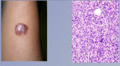
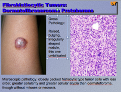
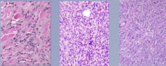
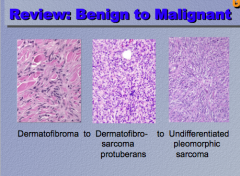
Raised, bulging, umbilicated
Less order in histiocyte cells, more cellularity and atypia than dermatofibroma.
Diagnosis? |
|
|
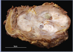
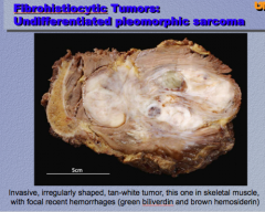
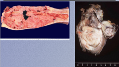
What is the disease? Focal hemorrhages, tan white tumor, skeletal muscle. |
|
|
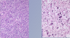
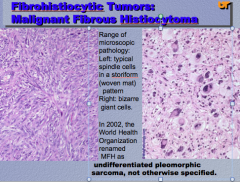
What cell types do you see? What is the pattern? What is the diagnosis? |
|
|
Identify the three.
Which is benign, which is malignant? |
|
|
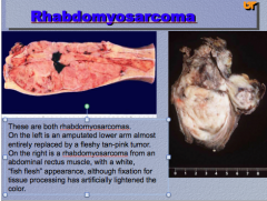
Fleshy-pink tumor, fish flesh appearance
What is the diagnosis? |
|
|
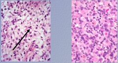
What type of cells shown? What type of cells recapitulate skeletal muscle cells? Diagnosis? |
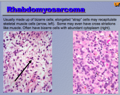
|
|
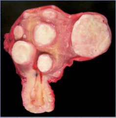
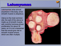
Where can this arise? Why?
What is the most common site? Why?
What is the diagnosis? |
|
|
What is the tumor? Describe. |
|
|
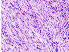
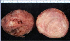
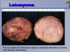
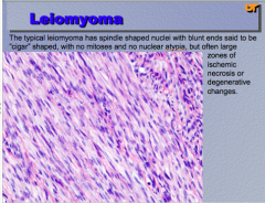
What do the cells look like? Are there mitoses and nuclear atypia? Large zones of what? Diagnosis? |
|
|
Diagnosis? |
|
|
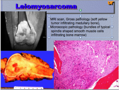
Leionmyosarcoma:
Which is low grade, high grade?
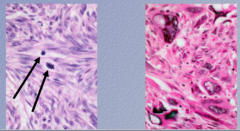
How do you recognize low grade as malignant? High grade? |

|
|
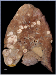
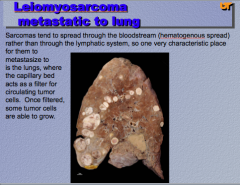
What is a characteristic place for leiomyosarcoma to metastasize? |
|
|
|
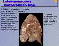
What is the major cause of death for patients with sarcomas?
In what part of the lung are metastases larger and more numerous? |
|