Reading...
![]()
Play button
![]()
Play button
![]()
Use LEFT and RIGHT arrow keys to navigate between flashcards;
Use UP and DOWN arrow keys to flip the card;
H to show hint;
A reads text to speech;
181 Cards in this Set
- Front
- Back
- 3rd side (hint)
|
Cornea
a. Epithelium on outer surface b. Name of outer basement membrane c. Name of inner basement membrane d. Type of internal epithelium |
a. Stratified squamous, nonkeratinized
b. Bowman's c. Descemet's d. Endothelium |
|
|
|
The 10 layers of the retina from internal (next to vitreous humor) to external
|
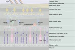
1. Inner limiting membrane
2. Optic nerve fiber 3. Ganglion cell layer 4. Inner plexiform layer 5. Inner nuclear layer 6. Outer plexiform layer 7. Outer nuclear layer 8. Outer limiting membrane 9. Photoreceptor cells 10. Pigment cells |
|
|
|
Nerves of orbit
a. CN III - Divisions and their innervation b. CN V1 - Branches and innervation |
a. CN III
I. Superior division - Levator palpebrae superioris, superior rectus II. Inferior division - The rest b. CN V1 I. Frontal nerve -> Supraorbital, supratrochlear II. Lacrimal nerve - Carries sensory and parasympathetic impulses to the lacrimal gland III. Nasociliary nerves - Infratrochlear nerve, long ciliary nerves, anterior and posterior ethmoidal nerves |
|
|
|
Ear
a. Reticular lamina - Function b. the hair cells are supported by c. Inner tunnel - Made by, function |
a. Prevents abnormal displacement of the stereocilia of hair cells and subsequent depolarization
(Rigid plate of cytoplasm) b. Phalangeal cells c. Inner tunnel I. Made by inner and outer pillar cells (Filled with microtubules to provide a rigid, triangular inner tunnel) II. Remains rigid to allow the organ of Corti to rock back and forth, instead of collapsing on itself by the vibrations |
|
|
|
Auricle
a. Ligaments b. Extrinsic muscles of auricle c. Proper muscles d. Motor innervation |
a. Lig. auriculare ant., sup., post.
b. Extrinsic muscles of auricle I. Mm. auriculares ant., post., sup. II. M. Temporoparietalis c. Proper muscles I. M. tragicus II. M. antitragicus III. Mm. Helicis major & minor IV. Mm. obliquus & transversus auriculae V. M. pyramidalis auriculae d. Facial nerve |
|
|
|
Auricle
a. Parts of helix (rim) b. Concha - Parts |
a. Helix
1. Crus 2. Spina 3. Cauda 4. Tuberculum Auriculare Darwini 5. Apex auriculae b. Cymba, cavitas |
|
|
|
Auricle
a. Arterial supply b. Venous drainage c. Lymphatic drainage d. Sensory innervation |
a. Temporal artery -> Anterior auricular branches
b. External jugular vein c. Parotid and mastoid lymph nodes d. Sensory innervation I. Auriculotemporal nerve -> Anterior auricular nerves II. CN VII -> Posterior auricular nerve III. CN X -> Auricular branch |
|
|
|
Meatus acusticus externus
a. Path b. Structure |
a. Path of external acoustic meatus
1. Opening\porus -> 2. Oblique ventromedial -> 3. Medial -> 4. Oblique ventromedial (= 160 degrees ventrally and convex downward) b. Structure I. Outer 2\3 - Cartilage (Lamina tragi - A longitudinal curved plate of cartilage, the beginning of the cartilaginous portion of the external acoustic meatus) II. Inner 1\3 - Bony, incisura tympanica Rivini\Tympanic notch (The notch in the superior part of the tympanic ring, bridged by the flaccid part of the tympanic membrane) III. Filled with 1. Tragi (The hairs growing at the entrance of the external acoustic meatus) 2. Glandulae ceruminosae (Tubuloalveolar glands, waxy cerumen, modified apocrine glands) |
|
|
|
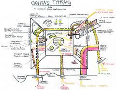
Tympanic cavity\Middle ear
a. Walls\paries and their components (Except for posterior wall) b. The 4 arteries and their origin |

a. Walls\Paries
1. Paries tegmentalis\Tegmental wall - Roof 2. Paries jugularis\Jugular wall - Floor I. Prominentia styloidea\Styloid prominence (Correspond to the base of the styloid process) 3. Paries labyrinthicus\Labyrinthine wall - Medial I. Promontorium (Rounded eminence, caused by the first coil of the cochlea) II. Sulcus promontorii\Groove of promontory (Branched groove running vertically, lodge tympanic plexus) III. Fenestra vestibuli\Oval window IV. Fenestrae cochlea\Round window (Covered by membrana tympanica secundaria) 4. Paries mastoideus\Mastoid wall - Posterior 5. Paries caroticus\Carotid wall - Anterior 6. Paries membranaceus\Membranous wall - Lateral b. Arterial supply tympanic cavity 1. Ascending pharyngeal artery -> fossula petrosa -> canaliculus tympanicus -> Inferior tympanic artery 2. External carotid -> Posterior auricular artery -> Foramen stylomastoideum -> Canaliculus chordae tympani posterior -> Posterior tympanic artery 3. Middle meningeal artery\A. meningea media -> Canalis nervi petrosi minoris -> Superior tympanic artery 4. Maxillary artery -> petryotympanic fissure -> Anterior canal of chorda tympani -> Anterior tympanic artery |
|
|
|
Paries mastoideus\Mastoid wall of tympanic cavity - Prominences
|
Paries mastoideus
1. Aditus ad antrum mastoideum 2. Prominentia canalis semicircularis lateralis 3. Prominentia canalis n VII 4. Eminentia pyramidalis (A conical projection to the vestibular window in the middle ear. it is hollow and contains the stapedius muscle) 5. Fossa incudis\Fossa of incus (A small depression in the lower and posterior part of the epitympanic recess that lodges the short limb of the incus) 6. Apertura tympanica canaliculi chordae tympani\Tympanic aperture of canaliculus for chorda tympani (Lateral to the pyramidal eminence) |
|
|
|
External acoustic meatus
a. Arterial supply b. Venous drainage c. Lymphatic drainage d. Innervation |
a. Arterial supply
I. Temporal artery -> Anterior auricular branches II. Maxillary artery ->deep auricular artery b. Venous drainage I. V. temporalis spf -> (+ maxillary vein = retromandibular vein) II. V. auricularis post. -> retromandibular vein c. Same as auricle - Parotid and mastoid lymph nodes d. Innervation I. CN X -> Auricular branch II. CN V -> Auriculotemporal nerve -> Branch of external acoustic meatus |
|
|
|
Tympanic membrane
a. Tympanic sulcus b. Size c. Fibrocartilaginous ring of tympanic membrane |
a. Tympanic sulcus
I. The groove on the inner aspect of the tympanic part of the temporal bone in which the tympanic membrane is fixed b. Size I. 0.1 mm thickness II. 9x10 mm c. Fibrocartilaginous ring of tympanic membrane\Gerlach annular tendon I. The thickened portion of the circumference of the tympanic membrane that is fixed in the tympanic sulcus |
|
|
|
Tympanic membrane
a. Umbo of tympanic membrane b. Malleolar stria c. Prominentia mallearis\Mallear prominence |
a. Umbo of tympanic membrane
I. The projection on the inner surface of the tympanic membrane at the end of the manubrium of the malleus II. Corresponds to the most depressed point of the membrane b. Malleolar stria I. A bright line seen through the membrana tympani II. Produced by the attachment of the manubrium of the malleus c. Mallear prominence I. A small prominence at the upper end of the stria mallearis produced by the lateral process of the malleus |
|
|
|
Membrana tympani
a. Mallear folds\Plicae mallearis b. Trigonum Woodi c. Pars flaccida - What, eponym d. Position e. Paracentesis - Where is it performed |
a. Mallear folds\Plicae mallearis
I. Ant. & post. II. Ligamentous bands III. Extend from each extremity of the tympanic notch to the mallear prominence IV. Mark the boundary between the tense and flaccid portions of the tympanic membrane b. Trigonum Woodi I. Site of reflection of light reflex of otoscope c. Pars flaccida of Shrapnell\Shrapnell membrane I. Triangular loose part of tympanic membrane between the malleolar folds c. Position I. Sagittal - 50 degrees declination II. Transvserse - 45 degrees inclination d. Lower posterior quadrant |
|
|
|
Membrana tympani
a. Arterial supply b. Lymphatic drainage c. Innervation |
a. Arterial supply
I. Maxillary artery -> a. auricularis prof. (Outer surface) II. Tympanic arteries (inner surface) b. Mastoid and parotid lymph nodes c. Innervation I. CN X -> Auricular branch II. CN V -> Auriculotemporal nerve -> branch of membrana tympani |
|
|
|
Middle ear
a. Arterial supply b. Venous drainage |
a. Arterial supply
I. 4 tympanic arteries (Anterior tympanic from maxillary) II. Caroticotympanic arteries (Of ICA) b. Venous drainage I. Plexus pterygoideus II. Sinus petrosus superior\Superior petrosal sinus |
|
|
|
Ossicles
a. Malleus - Parts b. Incus - Parts c. Stapes - Parts |
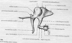
a. Malleus
1. Manubrium 2. Caput 3. Collum 4. Processus lateralis (Attached to membrana tympani) 5. Processus anterior (Slender spur running anteriorly from the neck of the malleus toward the petrotympanic fissure) b. Incus 1. Corpus 2. Crus longus\Processus lenticularis (Articulates with stapes) 3. Crus breve (The process that fits into a depression (fossa incudis) in the epitympanic recess) c. Stapes 1. Caput 2. Crus anterior and posterior 3. Basis\Foot plate |
|
|
|
Middle ear - Joints and type
|
Joints
1. Art. incudomallearis I. Sellar\Saddle 2. Art. Incudostapedia I. Spheric II. Between lenticular process of on the long crus of the incus and the head of the stapes 3. Syndesmosis tympanostapedialis |
|
|
|
Middle ear - Ligaments
|
Ligaments
1. Anterior ligament of malleus - 2 parts I. Meckel band - Pass from the base of the anterior process to the spine of the sphenoid through the petrotympanic fissure II. Anterior ligament of Helmholtz\Superior mallear ligament - From the anterior aspect of the neck of the malleus to the anterior boundary of the tympanic notch 2. Membrana stapedialis\Stapedial membrane I. The delicate mucosal layer that bridges the space between the crura and base of the stapes 3. L. anulare stapediale I. Ring of elastic fibers that attaches the base of the stapes to the margin of the fenestra vestibuli\oval window |
|
|
|
Antrum mastoideum
a. Antrum mastoideum b. Mastoid cells - Types c. Close relation to |
a. Antrum mastoideum\Mastoid antrum
I. A cavity in the petrous portion of the temporal bone II. Communicate posteriorly with the mastoid cells and anteriorly with the epitympanic recess of the middle ear via the aperture of the mastoid antrum b. Mastoid cells 1. Pneumatic type 2. Diploic type 3. Sclerotic type c. Sigmoid sinus |
|
|
|
Auditory tube
a. Synonyms b. Pharyngeal opening - Where, opened by which muscles |
a. Auditory tube\Tuba pharyngotympanica\Eustachian tube
b. Pharyngeal opening I. In the level of inferior nasal meatus II. Opened by tensor tympani and salpingopharyngeus (Contains glandulae tubariae) |
|
|
|
Inner ear\Auris interna
a. Bony labyrinth\Labyrinthus osseus - Parts b. Membranous labyrinth\Labyrinthus membranaceous - Parts |
a. Bony labyrinth
1. Vestibulum 2. Semicircular canals 3. Cochlea 4. Internal acoustic meatus 5. Perilymphatic space b. Membranous labyrinth 1. Labyrinthus vestibularis 2. Labyrinthus cochlearis 3. Spatium endolymphaticum |
|
|
|
Semicircular canals
a. Eminentia arcuata b. Crura - Which are common, which is singular |
a. Eminentia arcuata
I. Prominence on the anterior surface of the petrous portion of the temporal bone indicating the position of the superior semicircular canal b. Crus commune - Ant & post, crus simplex - Lateral |
|
|
|
Bony labyrinth - Cochlea
a. Cochlear cupula b. Basis cochlea c. Helicotrema |
a. Cochlear cupula
I. The domelike apex of the cochlea b. Basis cochlea\Base of cochlea I. The enlarged part of the cochlea that is directed posteriorly and medially and lies close to the internal acoustic meatus c. Helicotrema (spiral, trema - hole) I. A semilunar opening at the apex of the cochlea II. the scala vestibuli and scala tympani communicate via it |
|
|
|
Cochlea
a. Canalis spiralis cochlea b. Lamina spiralis c. Lamina spiralis - Components d. Hamulus of the spiral lamina |
a. Spiral canal of cochlea
I. The winding tube of the bony labyrinth that makes 2.5 turns about the modiolus of the cochlea II. Divided into two compartments by a winding shelf of bone - the bony spiral lamina b. Lamina spiralis ossea I. A double plate of bone winding spirally around the modiolus dividing the spiral canal of the cochlea incompletely into two - scala tympani and scal vestibuli II. Between the two plates of this lamina, the fibers of the cochlear nerve reach the spiral organ of Corti c. Lamina spiralis ossea 1. Lamella vestibularis I. The thicker of the two plates of bones II. The two plates are incompletely separated from each other by canals for peripheral fibers from the spiral ganglion III. Lies on the side of scala vestibuli IV. A thickening of the periosteum - the spiral limbus, is attached to the vestibular lamella within the cochlear duct 2. Lamella spiralis d. Hamulus of the spiral lamina I. The upper hooklike termination of the bony spiral lamina at the apex of the cochlea |
|
|
|
Cochlea
a. Cochlear canaliculus b. External opening of cochlear canaliculus |
a. Cochlear canaliculus
I. Minute canal in the temporal bone II. Passes from the cochlea inferiorly to open in front of the medial side of the jugular fossa III. It contains the perilymphatic duct c. External opening of cochlear canaliculus I. The external opening of the cochlear aqueduct on the temporal bone medial to the jugular fossa |
|
|
|
Modiolus
a. Modiolus b. Base of modiolus c. Lamina of modiolus d. Longitudinal canals of modiolus |
a. Modiolus
I. The central cone-shaped bone about which turns the spiral canal of the modiolus b. Basis modioli cochlea\Base of modiolus I. The part of the modiolus enclosed by the basal turn of the cochlea II. It faces the lateral end of the internal acoustic meatus c. Lamina of modiolus I. A bony plate II. Extend upward toward the cupula III. Form with the hamulus the helicotrema d. Longitudinal canals of modiolus I. Centrally placed channels that convey vessels and nerves to the apical turns of the cochlea |
|
|
|
Internal acoustic meatus - Parts
|
Internal acostic meatus
1. Porus\opening acusticus internus 2. Fundus meatus acusticus internus (Lateral end of the internal acoustic meatus, the wall of which is formed by the thin cribriform plate of bone separating the cochlea and vestibule from the internal acoustic meatus) 3. Crista transversa I. A horizontal ridge that divides the fundus into a superior and an inferior area II. In the superior area are the introitus of the facial canal and openings for the branches of the vestibular nerve to the utricle and to the ampullae of the anterior and lateral semicircular canals III. In the inferior part are openings for the cochlear nerve, and for branches of the vestibular nerve to the saccule and to the posterior semicircular canal 4. Crista verticalis 5. Area n VII 6. Area cochlearis - Tractus spiralis foraminosus I. The area inferior to the transverse crest II. Through which the filaments of the cochlear nerve pass to enter the cochlea III. Forms the base of the conical modiolus about which the cochlear canal spirals IV. Tractus spiralis foraminosus - Openings in the cochlear area of the bottom of the internal acoustic meatus through which the fibers of the cochlear nerve leave the bony labyrinth to enter the cranial cavity 7. Area vestibularis sup. + inf. 8. Foramen singulare I. Posterior to the cochlear area II. Transmits the nerves to the ampulla of the posterior semicircular duct |
|
|
|
Membranous labyrinth - Parts
|
Membranous labyrinth
1. Utricle - Macula, membrana statoconiorum 2. Sacculus - Macula, membrana statoconiorum 3. Semicircular ducts - Ampulla, crura, sulcus, cupula 4. Ductus utriculosaccularis 5. Ductus reuniens (Saccule->cochlear duct) |
|
|
|
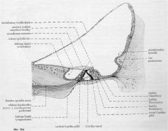
Membranous labyrinth - Labyrinthus cochlearis
a. Walls b. Spiral ligament c. Prominentia spiralis |

a. Walls
1. Paries vestibularis - Membrana vestibularis\Reissner membrane 2. Paries externus - Stria vascularis, prominentia spiralis, vas prominens, lig. spirale 3. Paries tympanicus - Crista basilaris, lamina basilaris, vas spirale b. Spiral ligament of cochlear duct\scala media I. The thickened periosteal lining of the bony cochlea II. Form the outer wall of the cochlear duct to which the basal lamina attaches c. Prominentia spiralis I. Projecting portion of the spiral ligament of the cochlea II. Bound to the lower edge of the stria vascularis III. Contain within a blood vessel - vas prominens |
|
|
|
Labyrinthus cochlearis
a. Crista basilaris b. Lamina basilaris |

a. Crista basilaris
I. Inward projection of the spiral ligament II. The basilar membrane\basal lamina of the cochlear duct is attached to it b. Basal lamina of cochlear duct I. The membrane extending from the bony spiral membrane to the basilar crest of the cochlea II. It forms the greater part of the floor of the cochlear duct separating the latter from the scala tympani III. Supports the organ of corti |
|
|
|
Labyrinthus cochlearis
a. Vas spirale b. Limbus spiralis c. Supporting cells of spiral organ |
a. Vas spirale
I. Large blood vessel that runs in the tympanic layer of the basilar membrane\basal lamina just beneath the tunnel of Corti (Corti tunnel - The spiral canal in the organ of Corti, formed by the outer and inner pillar cells\rods of Corti, filled with fluid) b. Limbus spiralis\Limbus of osseous spiral lamina I. The border of the spiral lamina II. The thickened periosteum covering the upper plate of the bony spiral lamina of the cochlea III. Labium limbi tympanici and labium limbi vestibularis c. Supporting cells of spiral organ of Corti 1. Pillars\Rods of Corti 2. Cells of Hensen 3. Cells of Claudius (The tectorial membrane is a gelatinous membrane that overlies the spiral organ of Corti) |
|
|
|
Endo- and perilymph
a. Endolymph - Composition, path b. Perilymph - Composition, path of drainage |
a. Endolymph
I. Similar to intracellular fluid II. Path - Stria vascularis -> scala media -> ductus endolymphaticus -> saccus endolymphaticus (blind) -> veins b. Perilymph I. Similar to ECF II. Drainage - Canalicus cochlea -> Subarachnoid space |
|
|
|
Inner ear - Nerves, ganglia, and their parts
|
1. Nervus vestibularis
I. Ggl vestibulare Scarpae II. Pars superior - N. utriculoampullaris III. Pars inferior - N saccularis, n ampullaris posterior 2. Nervus cochlearis I. Ggl cochleare Corti |
|
|
|
Association pathways\Fibrae associationis telencephali
a. What b. Short association pathways\Fibrae associationis breves b. Long association pathways\Fibrae assciationis longae |
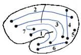
a. Connect regions in the same hemisphere
b. Short association pathways I. Fibrae arcuatae cerebri\Arcuate fibers of cerebrum b. Long association pathways 1. Fasciculus occipitofrontalis sup., inf (2,6) 2. Fasciculus longitudinalis sup., inf. (6,3) (Sup: Connect frontal, occipital, and temporal lobes. Frontal lobe -> operculum -> lateral sulcus -> occipital and temporal lobe) 3. Fasciculi occipitales verticales, horizontales (4,5) 4. Fasciculus uncinatus (1) (Frontal<-->Temporal lobes) 5. Cingulum (7) (Parahippocampal gyrus <--> Anterior perforated substance) |
|
|
|
Cortical commissural pathways\Fibrae commissurales telencephali - Which, fibers
|
Cortical commissural fibers
1. Fibrae corporis callosi I. Connect regions of all lobes II. Only auditory from the temporal lobe 2. Commissura fornicis I. Connects parahippocampal gyrus, dentate gyrus, and subiculum (Original commissure of archicortex) 3. Commissura anterior - Pars anterior I. Connects olfactory parts (Original commissure of paleocortex) 4. Commissura anterior - Pars posterior I. Connects regions of temporal lobe except auditory and hippocampal parts |
|
|
|
Commissural pathways of brain stem - Which, connections
|
Commissural pathways of brainstem
1. Commissura habenularum I. Connects habenular nuclei 2. Commissura posterior - Commissural fibers I. Connect posterior nuclei of thalamus, superior colliculi, and pretectal nuclei 3. Commissura posterior - Non-commissural fibers I. Fibers from interstitial nucleus of Cajal and posterior commissural nuclei of Darksevic to contralateral MLF |
|
|
|
Projection pathways - Ascending
a. Sensitive\Somatosensory b. Special sensory |
a. Sensitive
1. Direct I. Lemniscus system (lemniscus medialis) II. Anterolateral system (lemniscus spinalis) - Spinothalamic, spinoreticular, spinotectal, cranial nerves 2. Indirect - Cerebellar pathways b. Sensorial 1. Visual 2. Auditory 3. Vestibular 4. Olfactory 5. Gustatory |
|
|
|
Projection pathways - Descending
a. Pyramidal pathways b. Extrapyramidal pathways c. Descending pathways of the brainstem |
a. Pyramidal pathways
1. Corticospinal fibers 2. Corticonuclear fibers b. Extrapyramidal pathways 1. Tectospinal tract 2. Rubrospinal tract 3. Reticulospinal tract 4. Vestibulospinal tract c. Descending pathways of the brainstem 1. MLF 2. Fasciculus longitudinalis dorsalis Schutz |
|
|
|
Dorsal column-medial lemniscus system
a. Modalities b. Pathway |
a. Modalities - Touch, deep pressure, tension, vibrations, proprioception from joints, tendons, and muscle
b. Pathway 1st neuron I. Peripheral nerve II. Spinal ganglion III. Fasciculus gracilis et cuineatus IV. Synapse in nuclei gracilis et cuneatus 2nd neuron I. Internal arcuate fibers - decussate ('Decussatio lemnisci medialis) -> II. Lemniscus medialis III. Synapse in thalamus 3rd neuron I. Tractus thalamocorticalis -> II. Synapse in parietal lobe - postcentral gyrus area 3,1,2 (Tractus spino-bulbo-thalamo-corticalis, projection -> ascending -> sensitive -> direct, 3-neuronal pathway) |
|
|
|
Spinothalamic tract
a. Modalities b. Pathway |
a. Modalities
I. Sharp\fast pain, heat, cold, gross sensitivity b. Pathway (Type: projection -> ascending -> sensitive -> direct -> anterolateral system) |
|
|
|
Spinoreticular tract
a. Modalities b. Pathway |
a. Dull, "slow" pain. Activate ARAS
b. 2-neuronal pathway, end in reticular formation. Continue as tractus reticulothalamicus and other lines (Type: Projection -> Ascending -> Sensitive -> Direct -> Anterolateral system) |
|
|
|
Spinotectal tract
|
Spinotectal tract
I. Very old pathway II. Regressed into systems for motorics of muscles of eye, head, and neck |
|
|
|
Alternating hemiplegia
a. Alternating hemiplegia b. Hemiplegia alternans superior - What, eponym c. Hemiplegia alternans media - What, eponym d. Hemiplegia alternans inferior - What, eponym |
a. Alternating hemiplegia
I. Hemiplegia on one side with contralateral cranial nerve palsies b. Heimplegia alternans superior\Weber I. CN III c. Hemiplegia alternans media\Millard-Gubler I. CN VI d. Hemiplegia alternans inferior\Jackson II I. CN XII |
|
|
|
Tractus tectospinalis
a. Function b. Decussation |
a. Motorics of head and neck
b. Decussatio tegmenti posterior |
|
|
|
Tractus rubrospinalis
a. Function b. Decussation |
a. Function
I. Excitation of flexors and inhibition of extensors (Allow fine movement - extensors are more characterized by continuous activation for postural function) b. Decussatio tegmenti anterior |
|
|
|
Extrapryamidal motor systems
a. Tractus reticulospinalis - Function b. Tractus vestibulospinalis - Function |
a. Tractus reticulospinalis
I. Excitation of gamma-neurons II. Alternating excitation and inhibition of flexors and extensors b. Tractus vestibulospinalis I. Excitation of extensors (=Posture) |
|
|
|
Fasciculus longitudinalis medialis
a. Pathway b. Function |
a. Pathway
I. Uncrossed II. From 1. CN III 2. Superior colliculi 3. Interstitial nuclei of Cajal 4. Vestibular nuclei 5. Posterior commissural nucleus of Darksevitch III. To cervical spine b. Function I. Synkinesis of eyes, head and neck |
|
|
|
Fasciculus longitudinalis dorsalis
a. Eponym b. Pathway c. Function |
a. Schutz
b. Pathway I. Uncrossed II. Medial hypothalamus -> parasympathetic nuclei of CN, RF c. Function I. Bidirectional coordination of autonomic system |
|
|
|
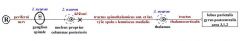
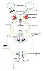
Optical pathway
|
Optical pathway
1st neurons - Rod and cones 2nd neurons - Bipolar cells 3rd neurons - Ganglionic cells 4th neurons - Lateral geniculate body (-> Geniculocortical tract\Radiatio optica Gratioleti -> Occipital lobe, area 17) |
|
|
|
Optical pathway - Branches from 3rd neuron\LGN
|
Optical pathway - Branches from 3rd neuron
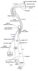
1. Retinohypothalamic tract I. Convert optical signals to the highest vegetative centers (See -> Salivate) II. Suprachiasmatic nuclei 2. Pathway of pupillary reflex I. Through area pretectalis 3. To Edinger-Westphal nucleus\Nucleus accessorius dorsalis CN III I. Miosis, accommodation 4. To reticular formation I. Tractus reticulospinalis -> centrum ciliospinale\Budge\C8-Th1 -> Sympathetic pathway in sympathetic trunk -> Superior cervical ganglion -> Plexus caroticus internus and ophthalmicus -> nn ciliares breves -> m dilator pupillae -> Mydriasis 5. Pathway for convergence I. -> Nucleus interstitialis Cajal -> MLF -> Oculomotor nuclei 6. Tectal optic circuit I. Tractus tectospinalis (Control of synkinesis of eye, head, neck to visual stimuli and for coordination with gross movements of body) |
|
|
|
|
|
|
|
Auditory pathway
|
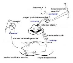
Auditory pathway - 4 neurons, crossed
1st neuron - Bipolar cell of cochlear ganglion -> I. Travel in CN VIII II. Synapse in cochlear nuclei 2nd neurons I. Cells in posterior cochlear nuclei -> striae medulares ventriculi quarti\medullary stria of fourth ventricle, dive into depth in median sulcus II. Cells of anterior cochlear nuclei - through trapezoid body III. Converge in lateral lemniscus IV. Synapse in inferior colliculi 3rd neurons - Cells in inferior colliculi 4th neurons - Cells of MGN I. -> Thalamus II. or -> Temporal lobe, transverse gyrus of Heschl, area 41 and 42 |
|
|
|
Vestibular pathway
|
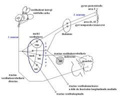
Vestibular pathway - 3 neurons, crossed
1st neuron I. Bipolar cells of vestibular ganglion II. CN VIII III. Synapse in vestibular nuclei (Part of fibers goes directly without relay as tractus vestibulocerebellaris directus) 2nd neuron I. Cells of nuclei vestibulares pontis II. Several pathways 1. Tractus vestibullaris indirectus to cerebellum 2. To spinal cord 3. To cranial nerve nuclei 4. To MLF 5. To RF 6. To thalamus 3rd neurons I. Cells of ventral nuclei of thalamis II. Synapse in temporal lobe, area 41,42, transverse temporal gyrus of Heschl and in area 3\postcentral gyrus of parietal lobe |
|
|
|
Olfactory pathway
|
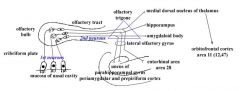
Olfactory pathway - 2 neurons
1st neuron I. Neuroepithelial cells in olfactory part of nasal cavity II. Fila olfactory\olfactory filaments cross through cribriform plate of ethmoid bone into cranial cavity 2nd neurons I. Mitral cells of olfactory bulb -> II. Olfactory tract -> III. Olfactory trigonum -> IV. Gyri\Stria olfactory med. et lat.-> 1. Limbic system - Amygdaloid body, temporal lobe: uncus gyri parahippocampalis, area entorhinalis\area 28, periamygdalar, and prepiriform cortex -> Hypothalamus, insular lobe 2. Nucleus mediodorsalis thalami -> orbitofrontal cortex\area 11 (+12,47?) |
|
|
|
Gustatory pathway
|
Gustatory pathway - 3 neurons
1st neuron - CN VII, IX, X 2nd neuron I. Nucleus tractus solitarius II. -> VPM of thalamus III. -> Motoric nuclei of cranial nerves IV. -> RF 3rd neuron - VPM I. -> Parietal lobe - area 43\gyrus postcentralis II. -> Rostral part of insula III. -> Parahippocampal gyrus IV. -> Habenula |
|
|
|
Sensory motor loop
|

Primary motor, somatosensory, premotor <--> Supplementary motor
1. -> Supplementary motor area -> 2. Putamen -> 3. Globus pallidus int. segment, substantia nigra -> 4. VL of thalamus -> Supplementary motor area... |
|
|
|
Oculomotor loop
|

Posterior parietal, prefrontal <--> Frontal eye field
1. -> Frontal eye field -> 2. Caudate nucleus -> 3. Globus pallidus int\med segment, substantia nigra -> 4. VL and DM nuclei of thalamus -> frontal eye field.. |
|
|
|
Association loop
|

Posterior parietal, premotor <--> Prefrontal
1. --> Prefrontal -> 2. Head of caudate -> 3. Globus pallidus medius, substantia nigra -> 4. VL and DM nuclei of thalamus -> prefrontal.. |
|
|
|
Limbic loop
|
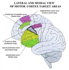
Medial and lateral temporal, hippocampal formation <--> ventral part of cingulate gyrus, orbitofrontal cortex
1. Ventral part of cingulate gyrus, orbitofrontal cortex -> 2. Ventral striatum, head of caudate -> (Ventral striatum - Nucleus accumbens, some nuclei of the olfactory tubercle) 3. Pallidum ventrale, globus pallidus medialis, substantia nigra -> 4. VA and DM nuclei of thalamus -> 1. |
|
|
|
Cerebellar output system - Ncl fastigii
|
Ncl fastigii ->
1. Lateral vestibular nucleus\Deiter 2. RF pontis and oblongata |
|
|
|
Cerebellar output - Interposed nuclei
|
Interposed nuclei (emboliform, globose) ->
1. Red nucleus 2. Thalamus 3. Inferior olive |
|
|
|
Cerebellar output - Dentate nuclei
|
Dentate nuclei ->
1. Lateral vestibular nuclei\Deiter 2. RF pontis 3. Superior olive 4. CN III 5. Interstitial nucleus of Cajal 6. Nuclei of Darkschewitz 7. Red nuclei |
|
|
|
Papez circuit
|
Papez circuit
1. Amygdala -> 2. Hippocampus -> 3. Fornix -> 4. Septum pellucidum -> 5. Mamillary bodies -> 6. Subthalamus -> 1. |
|
|
|
Direct vestibulocerebellar tract - Pathway
|
Direct vestibulocerebellar tract
1st neuron - Bipolar cell in vestibular ganglion 2nd neuron - granular cell in nodulus and flocculus (Function - Informations about head position in space and its movement. Transmits these info to eyeball muscles) |
|
|
|
Indirect vestibulocerebellar tract - Pathway
|
Indirect vestibulocerebellar tract
1st neuron - Bipolar cell in vestibular ganglion 2nd neuron - Cells in all vestibular nuclei 3rd neuron - Granular cells in nodulus, flocculus, and uvula (Function - Informations about head position in space and its movement. Transmits these info to eyeball muscles) |
|
|
|
Posterior spinocerebellar tract
a. Pathway b. Function |
a. Posterior spinocerebellar tract
1st neuron - Pseudounipolar cell in spinal ganglion 2nd neuron - Cells in Rexeds lamina V, VII -> Cerebellum b. Proprioception from lower limbs and lower half of the body |
|
|
|
Anterior spinocerebellar tract and cuneocerebellar tract
a. Function b. Path of cuneocerebellar tract |
a. Proprioception (kinetic part) from upper limbs
b. Cuneocerebellar tract 1st neuron - Pseudounipolar 2nd neuron - Nucleus cuneatus lateralis -> Mossy fibers |
|
|
|
Reticulocerebellar tract
a. Pathway |
a. Reticulocerebellar tract
1st neuron - T-cell 2nd neuron - VI Rexed lamina 3rd neuron - Precerebellar RF nuclei |
|
|
|
Pontocerebellar tract
a. Path b. Function |
a. Pontocerebellar tract
1st neuron - Cortical cell (premotor, motor (frontopontine), sensory (parietopontine), visual (tempero-occipito-pontine)) 2nd neuron - Pontine ncl b. Info about prepared movement |
|
|
|
Spino-olivo-cerebellar tract
a. Path b. Function |
a. Path
1. 1st neuron - T-cell 2. 2nd neuron - VI Rexed lamina 3. 3rd neuron - Accessory olivary nucleus -> Cerebellum b. Function I. Climbing fibers II. Integrative center for spino-cerebellar connections |
|
|
|
Cortico-olivo-cerebellar tract
a. Path b. Function |
a. Cortico-olivo-cerebellar tract
1st neuron - Area 4,6 2nd neuron - Inferior olive (Climbing fibers) b. Function I. Info about preceding movements |
|
|
|
Reticulocerebellar tract
a. Path b. Function |
a. Path
1st neuron - Motor, sensory, and visual cells 2nd neuron - Ncl reticularis potnis (Bechterew) - > Cerebellum b. Function I. Inform cerebellum about prepared movement under limbic system |
|
|
|
Cerebellar efferent tracts
|
Cerebellar efferent tracts
1. From deep cerebellar nuclei I. Brachium conjunctivum - Red nucleus, inferior olive, RF, VL & VA of thalamus II. Fasciculus uncinatus - RF, inferior olive, vestibular nuclei 2. From cerebellar cortex I. Tr. flocculo- et nodulo-vestibularis II. Tr. flocculo-reticularis III. Tr. Flocculo-nuclearis |
|
|
|
Archicortex - Parts
|
Archicortex
1. Gyrus dentatus 2. Cornu ammonis 3. subiculum 4. Indusium griseum 5. Area subcallosa 6. Stria longitudinales |
|
|
|
Periarchicortex - Parts
|
Periarchicortex
1. Area entorhinalis 2. Presubiculum 3. Parasubiculum 4. Area perirhinalis 5. Cingulate gyrus 6. Parahippocampal gyrus |
|
|
|
Limbic system 1-3 neurons
|
Limbic system
1. 1st neuron - Subiculum -> Fornix 2. 2nd neuron - I. Hypothalamus II. Mammillary bodies III. Anterior thalamic nuclei IV. Habenular nuclei V. Septal nuclei VI. Ncl accumbens VII. Amygdala 3rd neuron I. RF II. Entorhinal cortex III. Ncl interpeduncularis IV. Raphe dorsalis V. Nucleus accumbens VI. Amygdala |
|
|
|
Amygdalar connections - Subcortical
a. Stria terminalis b. Ventral amygdalo-fungal path c. Brainstem circuit |
a. Stria terminalis
1st neuron - Amygdala 2nd neuron - OF (olfactory?), ventromedial hypothalamic nuclei, area subcallosa, adolfactoria b. Ventral amygdalo-fungal path 1st neuron - Centromedial amygdala 2nd neuron - Anterior hypothalamic nuclei, ncl basalis, ventral striatum, medial hypothalamus c. Brainstem path 1st neuron - Centromedial amygdala 2nd neuron - Monoaminergic, RF, dorsal nucleus of CN X |
|
|
|
Auricle - Parts on posterior surface
|
Posterior surface
1. Eminentia conchae 2. Scaphae & fossa triangularis 3. Fossa antihelica 4. Sulcus crus helicis 5. Fissura antitragohelicina |
|
|
|
Middle ear
a. Lymph b. Innervation |
a. Lymph
I. Parotid lymph nodes II. Mastoid lymph nodes III. Deep cervical lymph nodes IV. Retropharyngeal lymph nodes b. Innervation I. CN IX -> N tympanicus II. V2 -> Pharyngeal branch (Inner part of auditory tube) III. Sympathetic - caroticotympanic nerves |
|
|
|
Auditory tube - Components of the wall
|
Components of wall
1. Pars cartilaginea I. Medial lamina of cartilage (The broad medial portion of the cartilaginous part) II. Lateral lamina of cartilage (The narrow lateral portion of the cartilaginous part) III. Membranous lamina of cartilage (The connective tissue membrane that, with the lateral and medial laminae, complete the lateral and inferior walls of the cartilaginous part of the pharyngotympanic tube) 2. Pars ossea I. Semicanal of auditory tube\Canal for pharyngotympanic tube (The inferior division of the musculotubal canal that forms the bony part of the pharyngotympanic tube) II. tubal air cells of pharyngotympanic tube\Cellulae pneumaticae (Occassional small air cells in the inferior wall of the pharyngotympanic tube, near the tympanic orifice, communicate with the tympanic cavity) |
|
|
|
Bulbus oculi - Parts
|
1. Tunica fibrosa - External
2. Tunica vasculosa - Media 3. Tunica interna - Nervosa 4. Lens |
|
|
|
Camerae bulbi - Parts
|
1. Anterior chamber
2. Posterior chamber 3. Vitreous chamber seu postrema |
|
|
|
Origin of the visual organ
|
1. Neuroectoderm of the forebrain
2. Surface ectoderm of the head 3. Mesoderm between 4. Neural crest cells |
|
|
|
Development of the visual organ
|
Development of the visual organ
I. Begin in the fourth week 1. Optic sulci in the neural folds -> 2. Evaginate into optic vesicles & optic stalks -> Optic cup (Optic vesicles - Evagination, > Retina. Optic stalk - Constricted proximal portion of optic vesicle, -> Optic nerve) (Optic cup - Double-walled cup, formed by invagination of the optic vesicle. Its inner layer becomes sensory retina, its outer layer the pigment retina) II. Induction of surface ectoderm 1. -> Lens placodes -> Lens pits -> Lens vesicles (Lens placodes - Ectoderma placodes\local thickening, Lens pits - Depression as lens placodes sink in toward the optic cup. Lens vesicle - Primordium of lens, ectodermal invagination) III. Vascular mesenchyme along optic stalk develops into vasa centralis retinae |
|
|
|
Origin of retina
|
1. Pigment epithelium <- Outer layer of optic vesicle
2. Inner layer <- Pars nervosa retinae (Is a intraretinal space that gradually disappears) |
|
|
|
Tunica nervosa - Retina - Parts
|
1. Pars caeca - Pars iridica, pars ciliaris
2. Pars optica - Discus n optici, macula lutea 3. Ora serrata |
|
|
|
Cones - photopigment
|
Iodopsin
|
|
|
|
Color vision - Spectra
|
1. Blue - 420 nm
2. Green - 535 nm 3. Red - 565 nm |
|
|
|
What forms external limiting membrane of retina
|
Muller cells (macroglial cells)
|
|
|
|
What forms internal limiting membrane of retina
|
Zonulae adherentes with rods and cones
|
|
|
|
Origin of the iris
|
From the anterior part\rim of the optic cup
1. Outer zone - epithelial cells -> smooth muscle 2. Inner layer - pigment epithelium |
|
|
|
Iris - Structure
|
1. Anterior surface - without epithelium
2. Posterior site I. Inner epithelial layer II. Outer myoepithelial layer 3. Stroma I. M sphincter pupillae II. M dilator pupillae |
|
|
|
Tunica vasculosa - Choroidea - Parts
|
1. Lamina suprachoroidea sclerae
(Loose, pigmented CT between the inner surface of the sclera and the outer surface of the choroid. The lamina fusca and suprachoroid lamina were considered to be two adjacent layers) 2. Lamina choriocapillaris\Capillary lamina of choroid (The internal or deep portion of the choroidea of the eye, composed fo a very close capillary network) 3. Lamina basalis choroidea Bruch (Transparent, nearly structureless inner layer of the choroid in contact with the pigmented layer of the retina) |
|
|
|
Cornea - Cells from
|
1. Surface ectoderm
2. Mesenchyme 3. Neural crest cells |
|
|
|
Choroid and sclera - Developed from
|
Adjacent mesenchyme
|
|
|
|
Tunica fibrosa - Sclera - Parts
|
1. Tenon capsule\Fascial sheath of eyeball
(Outer aspect of sclera, separated from sclera by episcleral space, attached to sclera near the sclerocorneal junction, blends with fascia of extraocular mm) 2. Lamina episclerais (Moveable loose CT, between the external surface of the sclera and tenon capsule) 3. Lamina suprachoroidea 4. Sclerocorneal junction\Angulus\Limbus corneae 5. Schlemm's canal (Fibroblasts + dense CT) |
|
|
|
Cornea
a. Vessels b. Layers |
a. None
b. Layers 1. Epithelium - Epithelium anterior cornea 2. Bowman membrane\lamina limitans anterior 3. Substantia propria cornea - Stroma 4. Descemet membrane - L. limitans posterior 5. Endothelium - Epithelium post corneae |
|
|
|
Lens development
|
Lens vesicle ->
(Ectodermal invagination that forms opposite the optic cup) 1. Anterior epithelium -> capsula lentis (thickened BM) 2. Posterior surface -> Primary fibers, secondary fibers |
|
|
|
Hyaloid artery
|
Terminal branch of primitive ophthalmic artery.
Forms vascular tunic around lens in vitreous body. (Atrophies almost completely by 8.5 months) |
|
|
|
The lens - Parts
|
1. Capsule - Elastic
2. Epithelium (anterior) - Simple, cuboidal 3. Matrix I. Cortical - Long cells (organelles, nuclei) II. Nuclear - Cell fibers (specific proteins - filensin, crystalins) 4. Zonula ciliaris Zinni\Suspensory ligaments of lens are elastic fibers that attach to the lens |
|
|
|
Conjunctiva
a. Epithelium b. Structure |
a. Stratified columnar
b. Structure 1. Glandulae conjunctivales Wolfringi 2. Caruncula lacramalis |
|
|
|
Eyelids - Development
|
1. 6th week - Surface ectoderm folds (containing mesenchyme) over the cornea
2. 10th week - Meet and adhere 3. Conjunctival sac forms meanwhile anterior to the cornea 4. 28th week - Eyes open |
|
|
|
Eyelids - Origin of muscles, tarsal plates, and glands
|
1. Muscles <- 2nd pharyngeal arch
2. Tarsal plate and glands <- Mesenchyme |
|
|
|
Eyelids\Palpebrae - Components
|
1. Sebaceous glands of Zeiss on outer skin surface
2. Ciliary glands of Moll on eyelashes 3. M orbicularis oculi 4. Tarsal plate - Tarsal\Meibomian glands (Fibroelastic plate, chalazion = chronic inflammatory granuloma of gland) |
|
|
|
Lacrimal gland - Origin, type
|
Surface ectoderm invaginations
(Out of order till the 6th week of postnatal life) Tubuloalveolar glands with myoepithelial cells. serous fluid (12-15 ducts lyzozyme-rich) |
|
|
|
Ear - origin
|
1. 1st pharyngeal pouch, groove, and membrane
2. 1st and 2nd pharyngeal arch mesenchyme 3. Surface ectoderm of the head 4. Neurofibers of the mesencephalon |
|
|
|
Auricle
a. Synonym b. Made of c. Origin |
a. Pinna
b. Elastic cartilage c. 2nd and 1st pharyngeal arches - auricular hillocks |
|
|
|
External auditory meatus - Origin
|
1st pharyngeal groove
(Groove is external) |
|
|
|
Tympanic membrane - Structure and origin
|
1. External covering - Very thin epidermis <- 1st pharyngeal groove
2. Tough collagen tissue <- 1st and 2nd pharyngeal arch mesoderm 3. Internal epithelium - Simple cuboidal <- 1st pharyngeal pouch |
|
|
|
Middle ear - origins
a. Tympanic cavity and tuba auditiva b. Auditory ossicles |
a. 1st pharyngeal pouch (Tubotympanic recess)
b. 1st and 2nd pharyngeal arches (Ossicles, tendons, and ligaments) |
|
|
|
Inner ear development
|
1. 4th week - Otic placode
(Otic placode - From surface ectoderm of the myelencephalon) - > Otic pits -> (Otic pits - Mark location of future auditory vesicles) 2. Otic vesicle\Otocyst -> Primordial labyrinth (Invaginated ectoderm, -> membranous labyrinth of inner ear) I. Dorsal utricular portion II. Ventral saccular portion 3. 20-22th week - Ossification of the cartilaginous otic capsule |
|
|
|
Basilar membrane - Thicker to ...
|
Apex
(Lower pitch sounds at apex) |
|
|
|
Cells of scala media
|
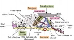
1. Inner and outer hair cells - have stereocilia
2. Supporting cells I. Pillar - Outer, inner II. Phalangeal III. Of Hensen IV. Of Claudius V. OF Boettcher |
|
|
|
Centers important for speech are
a. Area 44 b. Broca’s center c. Area 22 d. Area 39 e. Gyrus temporalis superior |
A,b,c,d,e
|
|
|
|
Optic pathway
a. Is 4-neuronal b. Terminates in area 17 c. Is conducted by tractus geniuclo-corticalis d. Is interpolated in colliculus superior e. Begins from primary receptor |
A,b,c,d,e
|
|
|
|
Auditory pathway
a. Is 4-neuronal b. Terminates in area 42 c. Terminates in gyrus temporalis transverses d. Is interpolated in inferior colliculus e. Is interpolated in corpus geniuculatum mediale |
A,b,c,d,e
|
|
|
|
Extrapyramidal tract
a. Is tractus spinoreticularis b. Is tractus spinorubralis c. Is tractus spinovestibularis d. Is tractus spinotectalis e. Conducts involuntary motor information |
E
|
|
|
|
To membranous labyrinth belong
a. Ductus reunions b. Ductus endolymphaticus c. Saccus endolymphaticus d. Ductus utriculosaccularis e. Ductus cochlearis |
A,b,c,d,e
|
|
|
|
Slow pain
a. Is interpreted in RF b. Is percepted in free nerve endings c. Is interpolated in ncl intralaminar thalami d. Has a cortical projection area 5 and 9 |
A,b,c,d
|
|
|
|
Somatosensory loop runs via
a. Area 4 b. Area 3,1,2 c. Putamen d. Globus pallidus e. Thalamus |
A,b,c,d,e
|
|
|
|
Fasciculus longitudinalis medialis
a. Begins in ncl interstitialis b. Begins in cajal’s nucleus c. Begins in ncl commissurae posterioris d. Begins in Darschevitsch nucleus e. Runs to vestibular nuclei |
A,b,c,d,e
|
|
|
|
Fasciculus longitudinalis posterior
a. Runs from hypothalamus b. Runs to RF c. Is decussated d. Runs to parasympathetic nuclei of CNs e. Is bidirectional |
A,b,d,e
|
|
|
|
To specific diencephalic nuclei belong
a. Ncl. Ventralis posteromedialis b. Ncl. ventralis posterolateralis c. Ncl. Corporis geniculati medialis d. Ncl. Corporis geniculati lateralis e. Ncl. Ventralis lateralis |
none
|
|
|
|
Fasciculus longitudinalis medialis
a. Begins in ncl interstitialis b. Begins in cajal’s nucleus c. Begins in ncl commissurae posterioris d. Begins in Darschevitsch nucleus e. Runs to vestibular nuclei |
A,b,c,d,e
|
|
|
|
Fasciculus longitudinalis posterior
a. Runs from hypothalamus b. Runs to RF c. Is decussated d. Runs to parasympathetic nuclei of CNs e. Is bidirectional |
A,b,d,e
|
|
|
|
Tractus pyramidalis
a. Begins from pyramidal cells b. Runs via crus posterius capsulae internae c. Runs via crus cerebri d. Begins from area 4 e. Runs via pyramids of medulla oblongata |
A,b,c,d,e
|
|
|
|
To hippocampal nuclei belong
a. Hippocampus b. Gyrus dentatus c. Subiculum d. Fornix e. Indusium griseum |
A,b,c
|
|
|
|
Which structures are to be found on the anterior surface of the middle ear cavity
a. Canaliculus caroticotympanici b. Canalis musculotubarus c. Fissure petrotympanica d. Eminentia pyramidalis e. Atticus |
A,b,c
|
|
|
|
Pain
a. Is conducted by tractus spinothalamicus anterior b. Is conducted by tractus spinoreticularis c. Is conducted by tractus spinobrachialis d. Is conducted by lemniscus spinalis e. Is suppressed by descending fibers from periaqueductal gray matter of mesencephalon |
A,b,c,d,e
|
|
|
|
Cerebellar nucleus
a. Is ncl dentatus series connected in neocerebellum b. Is ncl olivarius inferior c. Is ncl emboliformis series connected in archicerebellum d. Are ncll fastigi series connected in paleocerebellum e. Is ncl globosus series connected in archicerebellum |
none
|
|
|
|
Which layer belong to cornea
a. Squamous multilayered epithelium b. Flat one-layered epithelium c. Descemet membrane d. Lamina limitans anterior e. Epithelium anterius |
A,b,c,d,e
|
|
|
|
Tunica vasculosa bulbi (uvea) is composed of
a. Corpus ciliare b. Retina c. Iris d. Choroidea e. Corpus vitreum |
A, b,c
|
|
|
|
Medial wall of the orbit is composed of
a. Os ethmoidale b. Os lacrimale c. Ala major ossis sphenoidalis d. Maxilla e. Os zygomaticum |
A,b,d
|
|
|
|
Musculus obliquus superior
a. Inserts dorsally to the equator bulbi b. Passes across the spina trochlearis c. Is innervated by N IV d. Pulls the eyeball laterocaudally e. Begins above annulus tendineus communis |
A,b,c,d,e
|
|
|
|
To smooth muscles of optic apparatus belong
a. M ciliaris b. M tarsalis superior c. M tarsalis inferior d. M orbitalis superior e. M orbitalis inferior |
A,b,c
|
|
|
|
Structures responsible for tear drainage out of the eye are
a. Glandula b. Punctum c. Canaliculus d. Saccus e. Locus |
B,c,d
|
|
|
|
Humor aquosus
a. Springs from ciliary body b. Is absorbed in iris c. Is absorbed in iridocorneal angle d. Is absorbed in Schlemm’s canal e. Is absorbed in fontana’s spaces |
A,b,c,d,e
|
|
|
|
To optic apparatus glands belong
a. Glandulae sebaceae b. Glandulae tarsales c. Glandulae metatarsals d. Glandulae conjunctivales e. Glandulae lacrimalis |
A,b,d,e
|
|
|
|
Which structures opens into vestibulum
a. Ductus reunions b. Ductus endolymphaticus c. Canaliculus cochleae d. Crus commune e. Crus simplex |
D,e
|
|
|
|
Middle ear is supplied by branch of
a. Arteria pharynea ascendens b. Arteria auricularis posterior c. Arteria maxillaries d. Arteria meningea media e. Arteria carotis interna |
A,b,c,d,e
Arteria auricularis posterior -> Stylomastoid foramen -> anterior auricular artery |
|
|
|
Cavitas tympani forms cavities or pouches called
a. atticus\epitympanic recess b. b. recessus epitympanicus c. recessus hypotympanicus d. antrum mastoideum e. promontorium |
a,b,c,d
|
|
|
|
The wall of ductus cochlearis is
a. membrane vestibularis b. lamina basilaris c. membrane tectoria d. reissner’s membrane e. prominentia spiralis |
a,b,c,d,e
|
|
|
|
Middle ear is innervated by
a. chorda tympani b. n petrosus minor c. n petrosus major d. n VIII e. n VII |
b,e
|
|
|
|
Which structures are to be found in dermis
a. vater-paccini’s corpuscles b. meissner’s corpuscles c. merkel’s discs d. ruffini’s corpuscles e. langerhans’ corpuscles |
b, d
|
|
|
|
Olfactory mucosa
a. contains bowman’s glands b. contains primary receptors c. contains stem cells d. contains supporting cells e. contains neurons |
a,b,c,d,e
|
|
|
|
Taste buds are
a. primary ones b. situated close by the papillae vallatae c. situated in valleculae epiglotticae d. situated on the tongue root e. cleaned by serous ebner’s glands |
a,b,c,d,e
|
|
|
|
proprioception
a. is conducted by fasciculus cuneatus b. is felt by muscle spindles c. its cortical area is in the gyrus postcentralis d. is conducted via VPL\VPM nuclei e. is afflicted in syringomyelia |
a,b,c,d
|
|
|
|
Dorsal fascicule tracts (spino-bulbo-thalamic)
a. is decussated b. is 3-neuronal c. is conducted by lemniscus medialis d. conducts proprioceptive information e. conducts vibration information |
none
|
|
|
|
Cornea
a. is a derivative of neuroectoderm b. is supplied by the arteria ophtalmica c. is covered with conjunctiva d. its curved surface is a cause of the physiological astigmatism e. its internal surface is called the lamina limitans posterior |
b,c,d
(Astigmatism – A lens or optical system having different refractivity in different meridians. Meridian – A line encircling a globular body) |
|
|
|
Humor aquosus
a. is created in the zonula ciliaris b. is absorbed in the cloquet’s angle c. its higher pressure is a cause of the cataract d. its lower pressure is a cause of coloboma retinae e. is present in the camera postrema |
d
(Camera postrema\Postremal chamber of eyeball – the large space between the lens and the retina. Filled with vitreous body) (Coloboma – Any defect, congenital, pathological, or artificial, especially of the eye due to incomplete closure of the optic fissure) |
|
|
|
Retina
a. is a derivative of ectoderm b. its caecal part does not contain the cones c. its caceal part does not contain the rods d. its macula lutea is situated in the geometrical axis of the eyeball e. in avulsion retinae its pigmentous part adheres to the choroidea |
b,c,e
|
|
|
|
Lens
a. its fibers are produced by cells of its superficial neuroectodermal epithelium b. is supplied by the nervus hyaloideus during the fetal period c. is supplied via the lauth’s canal d. its capsule is a derivative of mesoderm e. its dimensions and shape changes depend on the contraction of the muscularis ciliaris |
e
|
|
|
|
Palpebra
a. hordeolum is an inflammation of its glandulae sebaceae b. contaisn the moll’s glands c. contains the tarsus d. is covered by both the skin and conjunctiva e. contains apocrine glands |
a,b,c,d,e
(Hordeolum – Suppurative inflammation of a gland of the eyelid) |
|
|
|
Position and movement of the
a. palpebra inferior is dependent on the nervus facialis b. palpebra superior is dependent on nervus facialis c. palpebra superior is dependent on nervus oculomotorius d. palpebra inferior is dependent on nervus oculomotorius e. palpebra superior si dependent on sympathetic |
a,c,e
|
|
|
|
Orbit contains following structures
a. tuberculum marginale b. processus orbitalis c. foramen nasolacrimale d. canalis supraorbitalis e. incisura frontalis |
a, b, e
(Incisura frontalis\frontal notch, orbital process of palatine bone) |
|
|
|
Which muscles pull the eye-ball laterally
a. musculus rectus bulbi lateralis b. musculus obliquus bulbi superior c. musculus obliquus bulbi inferior d. musculus orbicularis oculi e. musculus rectus bulbi medialis |
a,b,c
|
|
|
|
which structures is part of the auricle\pinna
a. cymba b. concha c. scapha d. tragus e. helix |
a,c,d,e
|
|
|
|
which embryonal layers are responsible for the tympanic membrane development
a. ectoderm b. mesoderm c. endoderm d. first branchial cleft e. first branchial pouch |
a,b,c,d,e
|
|
|
|
which structures is\are neighbors of the tympanic cavity
a. sinus sigmoideus b. vena jugularis interna c. arteria carotis interna d. sinus sphenoidalis e. lobus temporalis |
a,b,c,e
|
|
|
|
Recessus epitympanicus contains
a. corpus mallei b. caput incudis c. chorda tympani d. muscles stapedius e. basis stapedi |
c
|
|
|
|
which structures open into the utriculus
a. aquaeductus vestibule b. ductus reunions c. canalis semicircularis anterior d. canalis semicircular posterior e. canalis semicircularis lateralis |
none
|
|
|
|
Inner ear
a. helicotrema connects the scala tympani and scala vestibule b. canalis spiralis modioli contains the bipolar neurons c. canalis longitudinalis modioli contains the axons of bipolar neurons d. canaliculus cochlea contains the perilymph e. membrane tympani secundaria obturates the fenestra cochlea |
a,b,c,d,e
|
|
|
|
Sensory innervations of the acoustic apparatus is served by
a. V2 b. VII c. IX d. Plexus cervicalis e. V3 |
c,d,e
|
|
|
|
Inner ear is supplied by
a. Arteria labyrinthi b. Arteria cerebellaris anterior superior c. Arteria basilaris d. Arteria vestibulocochlearis e. Arteria carotis interna |
A,c, d
|
|
|
|
Gustatory buds
a. Contain stem cells b. Contain supporting cells c. Contain neuroepithelial cells d. Contain primary receptors e. Are present on the inferior surface of the tongue |
A,b,c
|
|
|
|
The olfactory organ
a. Is a derivative of the endoderm b. Is a pouch of the telencephalon c. Covers the concha nasalis superior d. Are cleaned by the ebner’s serous glands e. Its receptors are the multipolar neurons |
C
|
|
|
|
Epidermis contains
a. Meckel’s cells b. Langhans’s cells c. Free nerve endings d. Stratum germinativum e. Melanocyte epidermal unit |
C,d,e
|
|
|
|
Hypodermis contains
a. Vater-pacini corpuscles b. Stratum membranosum c. Scarpa’s fascia d. Colles’ fascia e. Corpuscula lamellosa |
A,b,c,d,e
(Corpuscula lamelloma\lamellated corpuscles\vater-pacini corpuscles) |
|
|
|
Which of the following people were real
a. Sandwich b. Boycotte c. Badminton d. Sartorius e. Mecenas |
A,b,c,e
|
|
|
|
Area pretectalis
a. Is a centre of the papillary reflex b. Is resposnibel for the coordination of the accommodation and convergence c. Is responsible for the accommodation reflex d. Is a centre of the circadial rhythms e. Contaisn the ncl perlia |
A,b,c,e
|
|
|
|
Autonomic system
a. Is controlled by the hypothalamus b. Is controlled by the RF c. Is controlled by the tractus reticulospinalis d. Its centres are connected via the MLF e. Its centres are connected via the tractus tegmentalis centralis |
A,b,e
|
|
|
|
Acoustic tract
a. Is interpolated in the thalamus b. Runs via the corpus trapezoideum c. Terminates in the lobus temporalis d. Its cortical centre is supplied by the ateria cerebri posterior e. Its second neuron is situated in the ncll cochleares (ant.post) |
a, B,c,e
|
|
|
|
Olfactory tract
a. Is 3-neuronal b. First neuron is situated in the bulbus olfactorius c. Is interpolated in the diencephalon d. Is interpolated in the ncl mediodorsalis e. Its cortical centre is supplied by arteria cerebri media |
none
|
|
|
|
Reticular formation
a. Dopamine is produced in the area ventralis tegmentalis tsai\ncl subbrachialis b. Dopamine is produced in the pars reticularis substantia nigra c. Serotonin is produced in the ncll raphes d. Acetylcholine is produced in the ncl basalis meynert e. Enkephalins is produced in the substantia grisea centralis |
A,c,d,e
(Substantia grisea centralis\Central gray substance – surrounding central canal of the spinal cord and the third and fourth ventricles) |
|
|
|
Cerebrospinal fluid is in the
a. Ventriculus lumbalis b. Cisterna sacralis c. Spatium subarachoideale d. Cavum septi pellucid e. Fissure telodiencephalica |
C
|
|
|
|
Control cerebellar circuits runs via
a. Nucleus ruber b. Ncll Olivares c. Ncll pontis d. Ncll VA + VL thalami e. Area 4 |
a,b
|
|
|
|
Papez circuit of the limbic system runs via
a. Epithalamus b. Tegmentum mesencephali c. Amygdale d. Fimbria hippocampi e. Corpus callosum |
D
|
|
|
|
Slow pain is transmitted via
a. Tractus spinothalamicus anterior b. Tractus spinothalamicus lateralis c. Anterolateral system d. Ncll VA, VL thalami e. Crus cerebri |
C
|
|
|
|
Corpus callosum connects
a. Lobus insularis b. Thalamus c. Hypothalamus d. Basal ganglia e. Hippocampal formation |
A
|
|
|
|
Voluntary movements
a. Are controlled in the area 5 b. The impulses run via the radiato thalami c. The impulses originate in the pyramidal cells of the fourth neocortical layer d. The impulses run via the tegmentum pontis |
none
|
|