Reading...
![]()
Play button
![]()
Play button
![]()
Use LEFT and RIGHT arrow keys to navigate between flashcards;
Use UP and DOWN arrow keys to flip the card;
H to show hint;
A reads text to speech;
82 Cards in this Set
- Front
- Back
- 3rd side (hint)
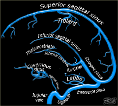
Superior Sagittal Sinus
|
occupies the attached convex margin of the falx cerebri. Commencing at the foramen cecum, the sinus is directed posteriorly, grooving the inner surface of the frontal bone, the adjacent part of the parietal bones and the inner aspect of the occipital bones. The SSS is triangular in cross section and gradually enlarges as it passes posteriorly.
|
|
|
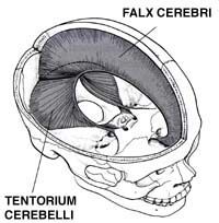
Falx Cerebri
|
Named because of its sickle-like shape, is a strong arched membrane that extends vertically downward in the longitudinal fissure between the two cerebral hemispheres.
|
|
|
|
Frontal Lobe
|
extends from the frontal pole of the brain to the central sulcus. Since it lies mostly in the anterior cranial fossa, it's lower surface is shallowly concave to fit the orbital roof. Some distance behind the frontal pole a prominent fissure, the lateral sulcus separates it from the temporal lobe.
|
The executive functions of the frontal lobes involve the ability to recognize future consequences resulting from current actions, to choose between good and bad actions (or better and best), override and suppress socially unacceptable responses, and determine similarities and differences between things or events.
The frontal lobes also play an important part in retaining longer term memories which are not task-based. These are often memories associated with emotions derived from input from the brain's limbic system. The frontal lobe modifies those emotions to generally fit socially acceptable norms. |
|
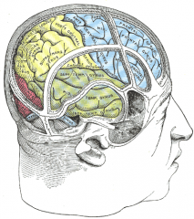
Parietal Lobe
|
lies between the frontal, temporal, and occipital lobes. It is separated from the frontal lobe by the central sulcus. Similarly, it is largely separated from the temporal lobe by the lateral sulcus, but at the end of the sulcus, parietal, temporal, and occipital lobes are confluent on the lateral surface of the hemisphere.
|
The parietal lobe plays important roles in integrating sensory information from various parts of the body, knowledge of numbers and their relations,[3] and in the manipulation of objects. Its function also includes processing information relating to the sense of touch.[4] Portions of the parietal lobe are involved with visuospatial processing.
|
|
|
Central Sulcus
|
is an important landmark because it forms the boundary between the frontal and parietal lobes and also separates the primary sensory cortex (posterior) from the primary motor cortex (anterior).
|
|
|
|
Superior Frontal Sulcus
|
separates the superior frontal gyrus from the middle frontal gyrus and may exist as a continuous sulcus or be divided into two or more parts (50%)
|
|
|
|
Precentral Gyrus
|
is bounded in front by the precentral sulcus and behind by the central sulcus. It serves as the primary motor region of the cerebral cortex also organized in a somatotropic fashion.
|
|
|
|
Postcentral Gyrus
|
lies nearly parallel to the precentral gyrus. It serves as the primary somesthetic cortex, in which general sensory projections are represented in a characteristic somatotropic pattern.
|
|
|
Cingulate Gyrus
|
is an arched convolution that lies in its entirety adjacent to the corpus callosum and is separated from it by the sulcus of the corpus callosum.
|
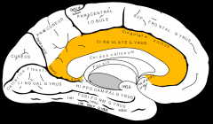
It receives inputs from the thalamus and the neocortex, and projects to the entorhinal cortex via the cingulum. It is an integral part of the limbic system, which is involved with emotion formation and processing, learning, and memory
|
|
|
Middle Frontal Gyrus
|
is a wide convolution that extends anteroinferiorly from the precentral gyrus and is bounded above by the superior frontal sulcus and below by the inferior frontal sulcus.
|
|
|
|
Superior Frontal Gyrus
|
is situated above the superior frontal sulcus and is a wide, uneven convolution continued on to the medial surface of the hemisphere.
|
|
|
|
Anterior Cerebral A.
|
is one of the terminal branches of the ICA. It is connected to the ACA of the opposite side by a short trunk called the anterior communicating artery.
|
|
|
|
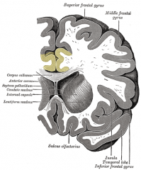
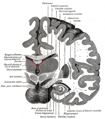
Lateral Ventricle
|
is an irregularly shaped cavity located within the lower and medial parts of each cerebral hemisphere, one on each side of the midline. The two ventricles are separated from one another by a thin median vertical partition, the septum pellicidum. Each communicates with the third ventricle and thus indirectly to each via the foramen of Monro.
|
|
|
|
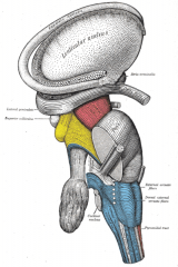
Caudate Nucleus
|
is an elongated mass of gray matter that is closely related to the lateral ventricle. Around the rostral border of the internal capsule, it is fused with the putamen of the lentiform nucleus while its tail terminates in close relationship to the amygdala.
|
|
|
|
Putamen
|
is the largest part of the basal ganglia and its most rostral part is located lateral to the head of the caudate nucleus, being separated by the anterior part of the internal capsule. It is the larger part of the lentiform nucleus which is comprised of the putamen and globus pallidus.
|
The main function of the putamen is to regulate movements and influence various types of learning. It employs GABA, acetylcholine, and enkephalin to perform its functions. The putamen also plays a role in degenerative neurological disorders, such as Parkinson's disease.
|
|
Genu/Splenium of Corpus Callosum
|
consists of fascicles of myelinated fibers whose main function appears to be the transmission of information between the neocortical portions of the two hemispheres during the learning process. The genu represents the curved anterior portion while the splenium is situated dorsal to the pineal body.
|
It connects the left and right cerebral hemispheres and facilitates interhemispheric communication. It is the largest white matter structure in the brain, consisting of 200–250 million contralateral axonal projections.
|
|
|
Middle Cerbral A
|
commences with the division of the ICA into its anterior and middle cerebral branches. Considered the largest branch, it appears as the lateral continuation of the ICA along the base of the brain above the lesser wing of the sphenoid bone. It supplies most of the frontal lobe, nearly all of the parietal lobe and most of the temporal lobe.
|
|
|
|
Frontal Horn of Lateral Ventricle
|
passes forward and laterally with a slight inclination downward beyond the interventricular foramen into the frontal lobe. Its rostral boundary is formed by the corpus callosum, medial by the septum pellucidum, and laterally by the head of the caudate.
|
|
|
Globus Pallidus
|
forms the more medial part of the lentiform nucleus and it is separated from the putamen by a thin layer of white matter called the lateral medullary lamina. It is considered the oldest part of the corpus striatum, and it receives its name from the fact that the many myelinated fibers that traverse its structure give it a lighter color than that of the putamen or caudate nucleus.
|
The globus pallidus is a structure in the brain which is involved in the regulation of voluntary movement. It is part of the basal ganglia, which, among many other things, regulate movements which occur on the subconscious level.
|
|
|
Foramen of Monro
|
permits communication of the third ventricle with each of the lateral ventricles on anterolateral aspect of the third ventricle.
|
|
|
|
Internal Capsule
|
is a thick mass of white fibers that bounded laterally by the lentiform nucleus, medially by the head of the caudate nucleus, the dorsal thalamus, and the tail of the caudate nucleus. Caudally, many of the fibers form the cerebral peduncles, while rostrally the fibers spread in a fan-like arrangement to become the corona radiata.. The anterior limb contains thalamocortical fibers, corticothalamic fibers, and frontopontine fibers. The genu additionally contains corticobulbar fibers to the motor nuclei of the cranial nerves. The posterior limb contains the central thalamic radiation which carries the thalamocortical somesthetic fibers to both the precentral and postcental gyri which communicate to the thalamus.
|
|
|
|
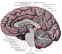
Third Ventricle
|
is a narrow, vertical, median cleft between the thalami of the two hemispheres. It communicates with the two lateral ventricles by the interventricular foramen. Caudally, it communicates with the fourth ventricle through the cerebral aqueduct.
|
|
|
|
Frontal Sinus
|
are situated behind the superciliary arches, are rarely symmetrical, and the septum between them deviates to one side or another of midline. Absent at birth, the frontal sinuses are fairly well-developed by the age of 7-8 years, but do not reach maximal size until after puberty.
|
|
|
|
Straight Sinus
|
is situated at the lone of the junction of the falx cerbri and the tentorium cerebelli. It is triangular in cross section, and it enlarges posterioly as it proceeds posteriorly from the end of the inferior sagittal sinus. It opens into the transverse sinus of the side opposite that in which the superior sagittal sinus terminates.
|
|
|
|
Vermis
|
lies between the cerebeller hemispheres and is continuous but unpaired median region. It's name is derived from the Latin word vermis meaning worm because of its shape.
|
|
|
|
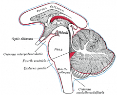
Optic Chiasma
|
is the convergence of the optic nerves in front and the divergence of the p[tic tracts behind. It rests upon the tuberculum sellae of the sphenoid bone. Lateral to the optic chiasma are the anterior perforated substance and the ICAs.
|
|
|
Insula
|
lies in the depths of the lateral sulcus on the lateral aspects of the cerebrum. The cortical gyri that overlap this structure are derived from the overgrowth of the frontal, parietal, and temporal lobes.
|
The insulae are believed to be involved in consciousness and play a role in diverse functions usually linked to emotion or the regulation of the body's homeostasis. These functions include perception, motor control, self-awareness, cognitive functioning, and interpersonal experience.
|
|
|
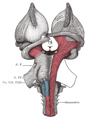
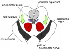
Midbrain
|
or mesencephalon is that part of the neuroaxis located between the pons inferiorly and the thalamus superiorly. It is a short, constricted segment of the brainstem that measures only about 2cm. By definition each half of the midbrain is called the cerebral peduncle, and this is further divided into a ventral part called the crus cerebri and a dorsal part called the midbrain tegmentum.
|
|
|
|
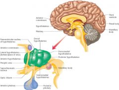
Mamillary Bodies
|
project prominently on each side of the ventral surface of the posterior hypothalamus near the midline
|
|
|
|
Posterior Cerebral A
|
in the adult commences as the terminal branches of the basilar artery. However, in about 20% it originates as a branch of the ICA. It supplies the areas of the brain responsible for visual motor behavior and visual perception.
|
|
|
|
Cerebral Aqueduct
|
connects the third and fourth ventricles; it has no choroid plexus. Blockage will result in hydrocephalus.
|
|
|
|
Cerebral Peduncle
|
is by definition half of the midbrain, also called the cerebral peduncle. This is further divided into a ventral part called the crus cerebri and a dorsal part called the midbrain tegmentum.
|
|
|
|
Optic Tract
|
leaves the chiasma and diverges from its fellow of the opposite side until it reaches the cerebral peduncle, where it winds obliquely across the undersurface. The fibers lying medially in the optic nerve cross in the chiasma and continue in the optic tract of the other side. The lateral fibers are uncrossed and continue to the brain in the optic tract of the same side.
|
|
|
|
Temporal Lobe
|
is the part of the hemisphere that lies below the lateral sulcus. The back end of this lobe is continuous with the occipital lobe.
|
|
|
|
Ethmoid Sinus
|
lie between the upper parts of the nasal cavities and the orbits, and are separated by these cavities by thin bony laminae. This sinus begins to develop after fetal life.
|
|
|
|
Middle Rectus
|
is one of the four recti that attaches to the bulb of the eye in such a manner that, acting singly, they turn its corneal surface in the direction corresponding to their names.
|
|
|
|
Cranial Nerve II
|
arises from the retinal ganglion cells. The central axons converge at the optic disc to form the optic nerve which enters the skull via the optic canal. Optic nerve axons terminate in the lateral geniculate body.
|
|
|
|
Lens
|
is a transparent, biconvex body; the convexity of its anterior surface is less than that of its posterior surface.
|
|
|
|
Internal Carotid A.
|
ascends from its origin in the neck posterolateral to the wall of the pharynx to enter the carotid canal in the lower surface of the petrous portion of the temporal bone.
|
|
|
|
Lateral Rectus
|
is one of the four recti that attaches to the bulb of the eye in such a manner that, acting singly, they turn its corneal surface in the direction corresponding to their names.
|
|
|
|
Pituitary Gland
|
lies immediately below the brain and is connected by a stalk, representing the stem by which the posterior lobe grew out from the brain. This gland is divided into two lobes each with its own tissue derivation and array of secretory hormones.
|
|
|
|
Dorsum Sellae
|
is composed of square plates of the sphenoid bone that end at superior angles in two turbucles as the posterior clinoid processes
|
|
|
|
Basilar A.
|
ascends along the basilar sulcus on the ventral aspect of the brain to the rostral border of the pons to divide into right and left posterior cerebral arteries.
|
|
|
|
Fourth Ventricle
|
is a flattened, diamond shaped cavity of the hindbrain and it contains the cerebrospinal fluid. It is situated ventral to the cerebellum and dorsal to the pons and upper half of the medulla. Its cavity is continuous with the cerebral aqueduct through the third ventricle. Inferiorly, its cavity opens below into the central canal, continuous with the spinal cord.
|
|
|
|
Cerebellum
|
is located dorsal to the pons and medulla and occupies the space between the brain stem and the occipital lobes of the cerebral cortex. It is connected to the brainstem by three peduncles. Although this structure probably does not initiate voluntary movement, it appears to serve as a supersegmental coordinator of muscular activity, especially those that require sequential, repetitive, or coordinated movements.
|
|
|
|
Uncus
|
is an anterior continuation of the lingual gyrus (temporal lobe) that is a hooklike, anterior structure partially covered by the periamygdaloid cortex.
|
|
|
|
Pontine Cistern
|
is a large space on the ventral aspect of the pons; within it courses the basilar artery. It is continuous caudally with the subarachnoid space and rostrally with the interpeduncular space.
|
|
|
|
Pons
|
is continuous above with the midbrain and inferiorly with the medulla oblongata
|
|
|
|
Nasal Septum
|
is bony in its large posterior component. The upper part of the bony septum is formed by the perpendicular plate of the ethmoid bone. Its lower part is formed by the vomer, with minimal contributions by the palantine and maxillary bones.
|
|
|
|
Sphenoid Sinus
|
is contained within the body of the sphenoid. Each sinus communicates with the sphenoethmoidal recess by means of an ostium in the upper part of its anterior wall. These sinuses are present as minute cavities at birth but maximal development occurs after puberty.
|
|
|
|
Clivus
|
is a shallow depression located posteriorly to the dorsum sellae. This structure supports the pons and the basilar artery.
|
|
|
|
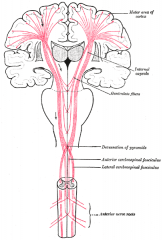
Medulla
|
is the direct upward continuation of the spinal cord through the foramen magnum which is itself continuous rostrally with the pons. The rearrangement of gray and white matter tracts more rostrally within the medulla, or bulb, as it is commonly known is such that at the level of the decussation of the pyramids, a transverse section of the brain stem is totally different from one through the spinal cord.
|
|
|
|
External Auditory Meatus
|
extends from the bottom of the concha to the tympanic membrance. It forms an s-shaped curve. It contains a cartilaginous portion and an osseous portion, which is slightly narrower and longer.
|
|
|
|
Mastoid Air Cells
|
are a hollowed out section of the mastoid process. The superior portion of this cavity is large and irregular, toward the inferior part it narrows and diminishes in size. Occasionally, these cells are completely absent.
|
|
|
|
Sigmoid Sinus
|
is directly continuous with the transverse sinuses and commences where the transverse sinuses emerge from the tentorium cerebelli. Each sigmoid sinus curves inferiorly and medially, in an s-shaped manner, in order to reach the posterior part of the jugular foramen where it terminates in the IJV.
|
|
|
|
Maxillary Sinus
|
is the largest of accessory sinuses of the nose. It is a pyramidal cavity located in the body of the maxilla. In the superior part of its medial wall is an ostium that communicates with the lower part of the infindibulum; a second or accessory orifice is frequently present in the middle meatus posterior to the first. This sinus appears as a shallow groove on the medial surface of the bone about the fourth month of fetal life, but does not reach its maximal size until after the second dentition.
|
|
|
|
Cerebellar Tonsil
|
are the lateral portions of the uvula of the cerebellum located in the posterior lobe.
|
|
|
|
Parotid Gland
|
is the largest of the 3 salivary glands. It lies upon the side of the face, immediately below and in front of the external ear.
|
|
|
|
Internal Jugular Vein
|
emerges deep to the posterior belly of the digastric muscle and receives one or more veins at the level of the hyoid bone. The one vein regularly entering the IJV at this point is the facial vein.
|
|
|
|
Mandible
|
the unpaired bone of the lower jaw consists of the tooth-bearing body and the more vertically disposed ramus that receives the insertions of the chief muscles of the jaw and articulates with the temporal bone. Ramus and body meet posteriorly at the angle of the mandible.
|
|
|
|
Spinal Cord
|
and its surrounding meninges lie in the vertebral canal. The space between the walls of the canal and the outer meninx of the cord, the dura mater, is the epidural space.
|
|
|
|
Soft Palate
|
is suspended from the posterior border of the hard palate. It consists of mucous membrane that encloses muscular fibers and vessels. When elevated, as in sucking and swallowing, it completely separates the nasopharynx from the oral cavity.
|
|
|
|
Tongue
|
is divided into lateral halves by a median fibrous septum, which extends its entire length and is fixed inferiorly to the hyoid bone. In either half there are two sets of muscles, extrinsic and intrinsic; the former have their origins outside the tongue, the latter within it. It is the primary organ of taste and an important organ of speech; it also assists in mastication and swallowing.
|
|
|
|
Uvula m.
|
arises from the posterior nasal spine of the palantine bones and from the palantine aponeurosis and inserts into the uvula.
|
|
|
|
Nasal Concha
|
are curved shelves of bone covered by mucosa that project from the lateral nasal wall and greatly increase the respiratory surface of the nose. Each concha is named according to its position with the inferior concha being longest. Above the superior concha may lie a subtle bulge referred to as the supreme concha.
a. Superior Concha b. Middle Concha c. Inferior Concha |
|
|
|
Sternocleidomastoid
|
extends obliquely upward and backward between the sternum and clavicle from each side of the neck to divide the cervical region of the neck into a anterior and lateral part. These parts form the anterior and posterior triangles.
|
|
|
|
Fornix
|
is a flattened fascicle of white matter which lies central to the septum pellucidum near the genu of the corpus callosum. It is the largest projection to the hypothalamus projecting from the hippocampal formation to the mamillary nucleus, anterior nucleus, and septal area.
|
|
|
|
Nasopharynx
|
lies posterior to the nose and above the level of the soft palate: it differs from the oral and laryngeal parts of the cavity in that is cavity always remains patent. It communicates with the choanae of the nasal cavities anteriorly.
|
|
|
|
Masseter m.
|
is a thick, quadralateral muscle composed of three parts which act to close the jaws by elevating the mandible. The fibers of the three parts blend and are continuous at their insertion on the ramus of the mandible.
|
|
|
|
Lateral Pterygoid muscle
|
is a short, thick muscle of two heads. It is somewhat conical in form and extends horizontally between the infratemporal fossa and the condyle of the mandible. The upper head arises from the infratemporal crest and the lateral surface of the greater wing of the sphenoid bone, while the inferior head arises from the lateral surface of the lateral pterygoid plate. It inserts on the neck of the condyle of the mandible and the articular disc of the TM joint.
|
|
|
|
Medial Pterygoid
|
is a thick, quadrilateral muscle that occupies the inner aspect of the ramus of the mandible, opposite of the masseter. It arises from the medial surface of the lateral pterygoid plate and the palantine bone. This muscle assists in closing the jaws by elevating the mandible. Acting together, they assist in protruding the mandible which assists in the chewing of food.
|
|
|
|
Thalamus
|
is the largest part of the diencephalon, but much of it is buried in the cerebral hemispheres. The thick right and left thalami are separated by the third ventricle.
|
|
|
|
Meningeal a.
|
give off branches to supply extracranial structures adjacent to it. They may enter the skull through the foramen ovale to reach the meninges around the trigeminal ganglion.
|
|
|
Amygdala
|
consists of a group of nuclei, located in the dorsomedial part of the rostral temporal lobe, that underlies or forms a bulge called the uncus.
|
processing of memory and emotional reactions, the amygdalae are considered part of the limbic system
|
|
|
Cranial Nerve I
|
arises in bipolar olfactory neurons and are responsible for smell. The central axons project to the olfactory bulb via the cribiform plate of the ethmoid bone.
|
|
|
|
Superior Rectus m.
|
is one of the four recti that attaches to the bulb of the eye in such a manner that, acting singly, they turn its corneal surface in the direction corresponding to their names.
|
|
|
|
Hypothalamus
|
region of the brain lying below the thalamus and making up the floor of the third cerebral ventricle. The hypothalamus is an integral part of the brain. It is a small cone-shaped structure that projects downward from the brain, ending in the pituitary (infundibular) stalk, a tubular connection to the pituitary gland. The hypothalamus contains a control centre for many functions of the autonomic nervous system, and it has effects on the endocrine system because of its complex interaction with the pituitary gland.
|
|
|
|
Red Nucleus
|
The red nucleus is a structure in the rostral midbrain involved in motor coordination. It comprises a caudal magnocellular and a rostral parvocellular part. It is located in the tegmentum of the midbrain next to the substantia nigra. The red nucleus and substantia nigra are subcortical centers of the extrapyramidal motor system
|
|
|
|
interpeduncular cistern
|
The interpeduncular cistern (basal cistern or Fossa interpeduncularis) is a wide cavity where the arachnoid extends across between the two temporal lobes.
It encloses the cerebral peduncles and the structures contained in the interpeduncular fossa, and contains the arterial circle of Willis. |
|
|
|
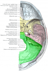
Posterior cranial fossa
|
The posterior cranial fossa is part of the intracranial cavity, located between the foramen magnum and tentorium cerebelli. It contains the brainstem and cerebellum.
This is the most inferior of the fossae. It houses the cerebellum, medulla and pons. Anteriorly it extends to the apex of the petrous temporal. Posteriorly it is enclosed by the occipital bone. Laterally portions of the squamous temporal and mastoid part of the temporal bone form its walls. |
|
|

Hippocampus
|
The hippocampus is located in the medial temporal lobe of the brain
It belongs to the limbic system and plays important roles in the consolidation of information from short-term memory to long-term memory and spatial navigation. In Alzheimer's disease, the hippocampus is one of the first regions of the brain to suffer damage; memory loss and disorientation are included among the early symptoms. Damage to the hippocampus can also result from oxygen starvation (hypoxia), encephalitis, or medial temporal lobe epilepsy |
|
|
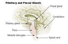
pineal gland
|
It produces the serotonin derivative melatonin, a hormone that affects the modulation of wake/sleep patterns and seasonal functions.[1][2] Its shape resembles a tiny pine cone (hence its name), and it is located near the centre of the brain, between the two hemispheres, tucked in a groove where the two rounded thalamic bodies join.
|
|