![]()
![]()
![]()
Use LEFT and RIGHT arrow keys to navigate between flashcards;
Use UP and DOWN arrow keys to flip the card;
H to show hint;
A reads text to speech;
100 Cards in this Set
- Front
- Back
|
What do epithelial tissue include? |
* Epithelia * Glands |
|
|
What are epithelia? |
Epithelia are layers of cells that cover internal and external surfaces
|
|
|
What are glands? |
Glands are structures that produce fluid secretions. |
|
|
What are the important characteristics of Epithelia? |
1) Cellularity 2) Popularity 3) Attachment 4) Avascularity 5) Regeneration |
|
|
What does the term "polarity' mean? |
The term polarity refers to the presence of structural and functional differences between the exposed and attached surfaces. |
|
|
What is attachment? |
The base of an epithelium is bound to a thin noncellular basement membrane which is also called the basal lamina. |
|
|
What are the functions of the Epithelial Tissue? |
* Provide Physical Protection * Control permeability * Provide sensation * Produce specialized secretion |
|
|
The epithelial cell is divided into two functional regions?
|
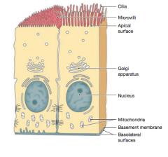
* Apical surface * Basolateral surfaces |
|
|
Identify the four major types of tissues in the body. |
1) Epithelial tissue 2) Connective tissue 3) Muscle tissue 4) Neural tissue |
|
|
What are Cell Adhesion Molecules (CAMs)? |
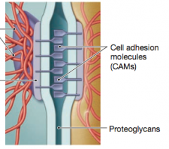
Cell Adhesion Molecules (CAMs) are proteins located on the cell surface involved in binding with other cells or with the extracellular matrix (ECM) in the process called cell adhesion. in essence, CAMs help cells stick to each other and to their surroundings. |
|
|
What are the three most common types of cell junctions?
|
* Tight Junctions * Gap junctions * Desmosomes |
|
|
What are gap junctions? |
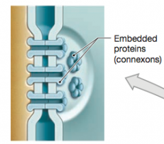
At a gap junction, two cells are held together by two interlocking transmembrane proteins called connexons. Two aligned connexons forms a narrow passageway that lets small molecules and ions pass from cell to cell. Gap junctions are common in cardiac muscle tissue and smooth muscle tissue because they are essential in coordinating muscle cell contractions. |
|
|
What are tight junctions? |
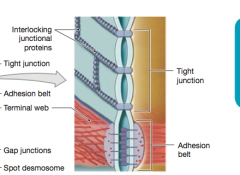
At tight junction, the lipid portions of the two plasma membranes are tightly bound together by interlocking membrane proteins. |
|
|
What are desmosomes? |
Desmosomes functions like rivets, fastening cells together into strong sheets. Intermediate filaments made of sturdy keratin proteins anchor desmosomes in the cytoplasm. Desmosomes attach muscle cells to each other in a muscle Some "muscle tears" involve the rupture of desmosomes. |
|
|
What is the probable functions of an epithelial surface whose cells bear many microvilli? |
An epithelium whose cells have many microvilli is probably involved in absorption or secretion. The microvilli greatly increase the surface area available for these processes. |
|
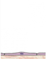
What type of tissue is this? Where can it be found? What are its functions? |
This is a Simple Squamous Epithelium. It can be found in the pleural, pericardial, and peritoneal cavities, as well as the lining of the heart and blood vessels. They reduce frictions, controls vessel permeability; performs absorption and secretion. |
|
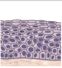
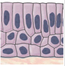
What type of tissue is this? Where can it be found? What are its functions? |
This is the Stratified Squamous Epithelium. They can be found in the surface of the skin, the lining of the mouth, throat, esophagus, rectum, anus, and vagina. They proceed physical protections against abrasion, pathogens, and chemical attack. |
|
|
The simple squamous epithelium that lines the body cavities is called__________. |
Mesothelium. |
|
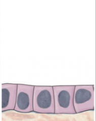
What type of tissue is this? Where can it be found? What are its functions? |
This is the Simple Cuboidal Epithelium. They can be found in glands, ducts; portions of kidney tubules; thyroid gland. They provide limited protection, secretion, and absorption. |
|
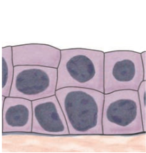
What type of tissue is this? Where can it be found? What are its functions? |
This is the Stratified Cuboidal Epithelium. They are found in the lining of some ducts but very RARE. They offer protection, secretion, and absorption. |
|
|
What is transitional epithelium and where can it be found? |
Transitional epithelium is an unusual stratisfied epithelium. Unlike most epithelia, it tolerates repeated cycles of stretching and recoiling without damage. A transitional epithelium is found in regions of the urinary system, such as the urinary bladder. |
|
What type of tissue is this? Where can it be found? What are its functions? |
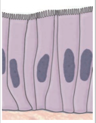
This is the Simple Columnar epithelium. They can be found in the lining of the stomach, intestine, gallbladder, uterine tubes, and collecting ducts of kidneys. They offer protection, secretion, and absorption. |
|
|
What is pseudo stratified columnar epithelium?
|
This is a columnar epithelium that includes several types of cells with varying shapes and functions. The distances between the cell nuclei and the exposed surface vary, so the epithelium appears to be layered, or stratified. However, it is not truly stratified, though, because every epithelial cell contacts the basement membrane. These cells also typically have cilia. *Epithelia of this type line most of the nasal cavity, the trachea (windpipe), the bronchi, and portions of the male reproductive tract. |
|
|
This is the Stratified Columnar Epithelium. This can be found in small area of the pharynx, epiglottis anus, mammary glands, salivary gland ducts, and urethra. They offer solely protection. |
|
|
What is endothelium? |
The simple squamous epithelium lining the inner surface of the heart and all blood vessels. |
|
|
What is the definition of a gland? |
A gland is a collection of epithelial cells that produce secretions. |
|
|
What are endocrine glands? |
Endocrine glands release their secretions into the interstitial fluid. These secretions are called hormones. Because endocrine gland secretions are not released into ducts, they are often called ductless glands. |
|
|
What are exocrine glands? |
Exocrine glands release their secretions into passageways called ducts that open onto an epithelial surface. Examples of exocrine secretions are enzymes entering the digestive tract, perspiration on the skin, tears in the eyes, and milk produced by mammary glands. |
|
|
What are the three modes of secretion? |
1) Merocrine secretion 2) Apocrine secretion 3) Holocrine secretion |
|
|
What is merocrine secretion? |
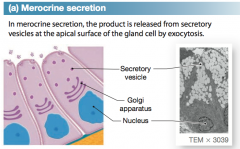
This is the most common mode of exocrine secretions. In merocrine secretion, the product is released from secretory vesicles by exocytosis. Mucin is a merocrine secretion that mixes with water to form mucus. |
|
|
What is apocrine secretion? |
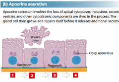
Apocrine secretion involves the loss of cytoplasm as well as the secretory product. |
|
|
What is holocrine secretion? |
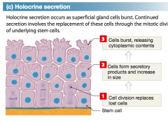
Holocrine secretion destroys the gland cell. During holocrine secretion, the entire cell becomes packed with secretary vesicles and then burst, releasing the secretion, but killing the cell. |
|
|
What are the ONLY unicellular exocrine glands in the body? |
The only unicellular exocrine glands in the body are mucous cells, which secrete mucins. Both the pseudo stratified ciliated columnar epithelium that lines the trachea and the columnar epithelium of the small and large intestines have an abundance of mucous cells. |
|
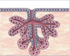
What is the structural classification of this gland? |
This is the simple tubular gland. They can be found in intestinal glands. |
|
What is the structural classification of this gland? |
This is the simple coiled tubular. They are the merocine sweat glands. |
|
What is the structural classification of this gland? |
This is the simple branched tubular. They are the gastric glands, the mucous glands of the esophagus, the tongue, and the duodenum |
|
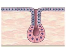
What is the structural classification of this gland? |
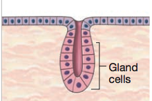
This is the simple alveolar (acinar). They are not found in adults; a stage in development of simple branched glands. |
|
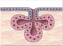
What is the structural classification of this gland? |
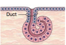
This is the simple branched alveolar. They are the sebaceous (oil) glands. |
|
What is the structural classification of this gland? |
This is the compound tubular. They are the mucous glands, bulbourethral glands (in the male reproductive system), and testes. |
|
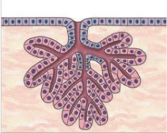
What is the structural classification of this gland? |
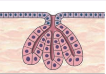
This is the compound alveolar (acinar). They are the mammary glands. |
|
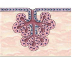
What is the structural classification of this gland? |
This is the compound tubuloalveolar. They are the salivary glands, the glands of respiratory passages, and the pancreas. |
|
|
Why do the pharynx, esophagus, anus, and vagina have similar epithelial organization? |
All these regions are subject to mechanical trauma and abrasion - by food (pharynx and esophagus), by feces (anus), and by intercourse or childbirth (vagina). |
|
|
What are the specific functions of the connective tissue? |
* Establishing a structural framework for the body * Transporting fluids and dissolved materials * Protecting delicate organs * Supporting surrounding, and interconnecting other types of tissue * Storing energy, especially in the form of triglycerides * Defending the body from invading microorganisms. |
|
|
What are the three classifications of connective tissues? |
* Connective tissue proper * Fluid connective tissues * Supporting connective tissues |
|
|
What is connective tissue proper? |
Connective tissue proper includes those connective tissues with many types of cells and extracellular fibers in a syrupy ground substance. Both adipose tissue (fat) [a loose connective tissue] and tendons [a dense connective tissue] are connective tissue proper, but they have very different structural and functional characteristics. |
|
|
What is fluid connective tissues? |
Fluid connective tissues have distinctive populations of cells suspended in a watery matrix that contains dissolved proteins. The only two types are blood and lymph. |
|
|
What is supporting connective tissues? |
Supporting connective tissues different from connective tissue proper in having a less diverse cell population and a matrix containing much more densely packed fibers. They two types of supporting connective tissues are cartilage and bone. |
|
|
In connective tissue proper, there are some cells that function in local maintenance, repair, and energy storage. What are they? |
They are * Fibroblasts * Fibrocytes * Adipocytes * Mesenchymal Cells These are permanent residents of the connective tissue. |
|
|
In connective tissue proper, there are some cells that defend and repair damaged tissues. They are more mobile. What are they? |
They are * Macrophages * Mast cells * lymphocytes * Plasma Cells * Microphages These are *not* permanent residents; instead they migrate through healthy connective tissues and collect at sites of tissue injury. |
|
|
What are fibroblasts? |
Fibroblasts are the only cells that are always present in connective tissue proper. They are also the most abundant permanent, or fixed, residents of connective tissue proper. Fibroblasts secrete hyaluronan and proteins. Each fibroblast also secretes protein subunits that assemble to form large extracellular fibers. |
|
|
What are fibrocytes?
|
Fibrocytes are the second most abundant fixed cell in connective tissue proper. They differentiate from fibroblasts. They maintain the connective tissue fibers of connective tissue proper. |
|
|
What are adipocytes? |
Adipocytes are also known as fat cells. |
|
|
What are mesenchymal cells? |
They are stems cells that are present in many connective tissues. These cells response to local injury or infections by dividing to produce daughter cells that differentiate into fibroblasts, macrophages, or other connective tissue cells. |
|
|
What are macrophages? |
They are large phagocytic cells scattered throughout the matrix. These scavengers engulf damaged cells or pathogens that enter the tissue. |
|
|
What are mast cells? |
They are small, mobile connective tissue cells that are common near blood vessels. The cytoplasm of mast cell is filled with granules containing histamine and heparin. |
|
|
What are lymphocytes? |
Lymphocytes migrate throughout the body, traveling throughout connective tissues and other tissues. Some lymphocytes may develop into plasma cells, which produce antibodies.
|
|
|
What are microphages? |
They are phagocytic blood cells that normally move through connective tissues in small numbers. They are attracted to the site of an infection or injury by chemicals released by macrophages and mast cells. |
|
|
What are melanocytes? |
They synthesize and store the brown pigment melanin, which gives tissues a dark color. They are abundant in connective tissues of the eye and the dermis of the skin. |
|
|
What the three types of fibers that occur in connective tissue? |
* Collagen * Reticular * Elastic Fibroblasts form all three by secreting protein subunits that interact in the matrix. |
|
|
What are collagen fibers? |
They are the most common fibers in connective tissue proper. They are long, straight, and unbranched. They are flexible like a rope, but it is stronger than steel when pulled from either end. Tendons which connect skeletal muscles to bones, consist almost entirely of collagen fibers. Typical ligaments are similar to tendons, but they connect one home to another bone. |
|
|
What are reticular fibers? |
Reticular ribers contain the same protein subunits as do collagen fibers, reticular fibers form a branching, interwoven framework that is tough, yet flexible. Reticular fibers stabilize the positions of an organ's blood vessels, nerves, and other structures, despite changing positions and the pull of gravity. |
|
|
What are elastic fibers? |
Elastic fibers are branched and wavy. After stretching, they return to their original length. |
|
|
What is ground substance? |
Ground substance fills the spaces between cells and surrounds connective tissue fibers. |
|
|
What is the loose connective tissue found in many parts of the embryo, including the umbilical cord? |
Mucous connective tissue |
|
|
What is loose connective tissue? |
Loose connective tissues are the packing materials of the body. They fill the spaces between organs, cushion, and stabilize specialized cells in many organs, and support epithelia. These tissue surround and support blood vessels and nerves, store lipids, and provide a route for the diffusion of materials. |
|
|
What are areolar tissue? |
Areolar tissue is the least specialized connective tissue in adults. Areolar tissue forms a layer that separates the skin from the deep structures. They cushion organs; provides support, but permits independent movement; phagocytic cells provide defense against pathogens. |
|
|
What is adipose tissue? |
They are fat. They are deep to the skin, especially at sides, buttocks, and breasts; padding around eyes and kidneys. They provide padding and cushion shocks; insulates; stores energy. |
|
|
What are reticular tissue? |
They are reticular fibers that form a complex three-dimensional storm. The storm supports the functional cells, or parenchyma, of these organs. They are found in the over, kidney, spleen, lymph nodes, and bone marrow. |
|
|
What is elastic tissue? |
Elastic tissue is a dense regular connective tissue made up mainly of elastic fibers. They help stabilize the positions of the vertebrae of the spinal column. |
|
|
In blood, the watery matrix is called _______. |
Plasm |
|
|
What type of connective tissue contains primarily triglycerides? |
Adipose (fat). |
|
|
Which two types of connective tissue have a fluid matrix? |
Blood and lymph |
|
|
Cartilage grows by two mechanisms, name them. |
* interstitial growth enlarges the cartilage from within * appositional growth adds new layers of cartilage to the surface. |
|
|
What are the three main types of cartilage? |
* Hyaline cartilage * Elastic cartilage * Fibrocartilage |
|
|
What hyaline cartilage? |
Hyaline cartilage is the most common type of cartilage. It is tough, but somewhat flexible because its matrix contains closely packed collagen fibers. They lie between the tips of ribs and bones of the sternum; covering bone surfaces at synovial joints; supporting larynx (voice box), trachea, and bronchi; forming part of nasal septum . They provide tiff but somewhat flexible support; reduce friction between bony surfaces. |
|
|
What is elastic cartilage? |
Elastic cartilage is extremely resilient and flexible because it contains numerous elastic fibers. They are found in the auricle of external ear; epiglottis; auditory canal; cuneiform cartilage of larynx. They provide support, but tolerates distortion without damage and returns original shape. |
|
|
What is fibrocartilage? |
Fibrocartilage is extremely durable and tough because it has little ground substance and its matrix is dominated by densely interwoven collagen fibers. They pad the knee joint; between pubic bones of pelvis; intervertebral discs. They resist compression; prevents bone to bone contact; limits movement. |
|
|
Osseous tissues are what?
|
Bone |
|
|
What are the four types of membranes found in the body? |
1) Mucous membranes 2) Serous Membranes 3) Cutaneous membrane 4) Synovial membranes |
|
|
What is mucous membranes? |
Mucous membranes line passageways and chambers that communicate with the exterior, including those in the digestive, respiratory, reproductive, and urinary tracts. |
|
|
What is the cutaneous membrane? |
The cutaneous membrane is the skin that covers the surface of your body. It consists of a *stratified squamous epithelium* and a layer of areolar tissue reinforced by underlying dense irregular connective tissue. |
|
|
What is the synovial membrane? |
The synovial membrane lines the joint cavity with synovial fluid. The epithelium has gaps and develops within a connective tissue. |
|
|
What are fasciae? |
Fasciae are connective tissue layers and wrappings that support and surround organs. When can divide the fasciae into three types of layers: * The superficial fascia * The deep fascia * The subserous fascia |
|
|
What is the superficial fascia? |

The superficial fascia is also called the hypodermis or subcutaneous tissue. This layer of areolar tissue and fat separates the skin from underlying tissues and organs. It proves insulation and padding, and lets the skin and underlying structures move independently. |
|
|
What is the deep fascia? |
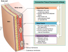
The deep fascia consists of dense irregular connective tissue. The organization of the fibers is like that of plywood: in each layer all the fibers run in the same direction, but the orientation of the fibers changes from layer to layer. |
|
|
What is the subserous facia? |

The subserous fascia is a layer of areolar tissue that lies between the deep fascia and the serious membranes that line body cavities. |
|
|
What are the three types of muscle tissue? |
1) Skeletal muscle, which forms the large muscles that produce gross body movements 2) Cardiac muscle, found in the heart, is responsible for circulating the blood 3) Smooth muscle, found in the walls of visceral organs and a variety of tore locations, where it provides elasticity, contractility, and support. |
|
|
What is the neural tissue? |
This is also known at the nervous tissue or nerve tissue. It is specialized for the propagation of electrical impulses from one region of the body to another. |
|
|
What are neuroglia? |
They are supporting cells for the neuron cells. |
|
|
What is inflammatory response? |
Inflammatory response isolates the injured area while damaged cells, tissue components, and any dangerous microorganisms, which could cause infection are cleaned up. |
|
|
What is regeneration? |
Regernation is the repair process that restores normal function after inflammation has subsided. |
|
|
The most abundant connections between cells in the superficial layers of the skin are________. |
Desmosomes. |
|
|
What are the three basic components of connective tissue? |
Connective tissues contain (1) specialized cells (2) extracellular protein fibers (3) a fluid ground substance |
|
|
What is the basement membrane? |
The basement membrane, also called the basal lamina, is a complex structure produced by the basal surface of the epithelium and the underlying connective tissue. |
|
|
A layer of glycoproteins and a network of fine protein filaments that prevents the movement of proteins and other large molecules from the connective tissue to the epithelium describes the ________.
|
The basement epithelium. |
|
|
Which type of muscle tissue is voluntary and contains large, multinucleate cells that can be up to 1 foot in length? |
Skeletal. |
|
|
What are the muscle tissues that produce movement of the body, movement of blood within the cardiovascular system, and movement of foodstuffs within the digestive tract? |
* Skeletal * Cardiac * Smooth Muscle |
|
|
What are the muscle tissues that are categorized as involuntary? |
* Cardiac * Smooth |
|
|
What are the muscle tissues that can regenerate after an injury? |
* Smooth |
|
|
What muscle tissue has extensive connections between cells at intercalated discs? |
Cardiac muscle |