![]()
![]()
![]()
Use LEFT and RIGHT arrow keys to navigate between flashcards;
Use UP and DOWN arrow keys to flip the card;
H to show hint;
A reads text to speech;
40 Cards in this Set
- Front
- Back
|
Cervical ribs~ supernumerary rib |
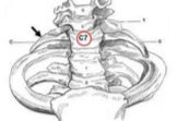
Associated with Thoracic Outlet Syndrome Compressed neurovasculature exiting Superior Thoracic Aperture ~0.5% - 2% population have a cervical rib Leads to confusion of vertebral levels in diagnostic images |
|
|
Lumbar ribs ~supernumerary rib |
Rarely associated with any clinical condition Articulates with ventral surface of the lumbar transverse processes <1% of population Misdiagnosed as Fx of L1 transverse process |
|
|
Flail chest |
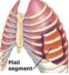
Life-threatening and unstable injury of thoracic wall Multiple rib fractures leads to detachment of thoracic wall to rib cage Intra-pleural pressure leads to paradoxical motion in breathing cycle |
|
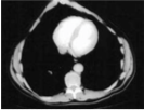
Pectus excavatum "sunken chest" |
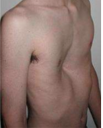
Concave depression Congenital thoracic wall deformity: intrauterine pressure on the chest wall during development Compression of heart and lungs |
|
Pectus carinatum "pigeon chest" |
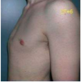
Protrusion of sternum and costal cartilages Connective tissue disorder Congenital thoracic wall deformity: Scoliosis and congenital heart disease
|
|
|
Sternal fracture |
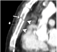
Injury to: 1. superior mediastinal structures 2. heart 3. lungs 4. major vessels |
|
|
Xiphoid process fracture |
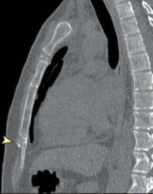
Xiphisternal joint dislocation Injury to: 1. Diaphragm Broken pieces can puncture: 2. Heart 3. Liver Poor CPR/contact sports |
|
|
Xiphisternal joint demarcates what? |
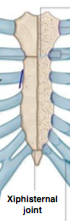
1. inferior limit of heart 2. central thoracic cavity 3. superior limit of liver 4. anterior diaphragm 5. Rib 7 attachment site 6. T10 vertebral body 7. T6 dermatome - xiphoid |
|
|
Weakest part of rib? |
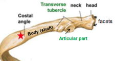
Most fractures occur in anterior costal angle |
|
|
"Slipping rib" syndrome |
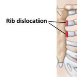
Rib dislocation sternocostal joint displacement Injury to diaphragm, liver, neurovasculature |
|
|
Rib separation |
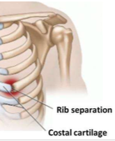
costochondral joint dislocation Separated rib may be displaced superiorly and often overrides the rib above |
|
|
Active inspiratory and expiratory accessory muscles? |
Active inspiration muscles: 1. sternocleidomastoid, 2. scalene muscle, 3. serratus anterior, 4. pectoral muscle Active expiration muscles: 1. internal intercostal, 2. innermost intercostal, 3. abdomen intercostal |
|
|
Severe dyspnea |
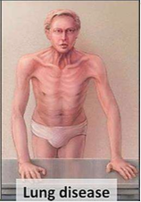
Recruitment of accessory muscles of respiration in shortness of breath 1. strenuous activity 2. lung disease (you can see his sternocleidomastoid in use) |
|
|
Passive respiratory muscles? |
Inspiration: 1. Diaphragm mainly and 2. external intercostals Expiration: elastic recoil in lungs (no muscles) |
|
|
Intercostal nerve block |
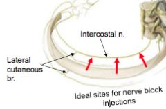
Nerve anesthetic 1. proximal mid-axillary line (same as chest tube) 2. Just before lateral cutaneous branches come off |
|
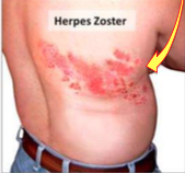
Herpes zoster |
Reactivation of chicken pox viruses which reside in Dorsal root ganglion Segmental innervation of thoracic wall by intercostal nerves Sx: sharp, burning pain, skin eruption in the strip of a dermatome |
|
|
Coronary Bypass Graft/Surgery |
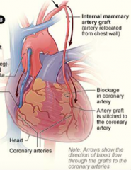
Coronary arteries which supply the heart can be blocked and a vessel we can choose to bypass the artery is the internal thoracic artery Remove a portion of internal thoracic artery to bypass coronary artery |
|
|
Posterior intercostal veins drain into? |
Azygous/Hemizygous system T5-T12 |
|
|
Anterior intercostal veins drain into? |
Internal thoracic vein |
|
|
Internal thoracic veins follow? |
Internal thoracic arteries |
|
|
Rule of veins? |
Any vein that follows an artery that branches off Subclavian Artery drains into Brachiocephalic vein!! ex) internal thoracic veins because they follow internal thoracic arteries which is a branch of subclavian |
|
|
What are the 4 Parietal pleura? |
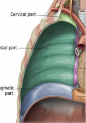
1. Cervical/Cupula 2. Costal 3. Mediastinal 4. Diaphragm |
|
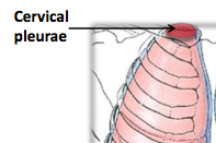
Cervical pleura (cupula) |
lung apex coming out of superior thoracic aperture 1st rib slopes inferiorly exposing pleura pleural site prone to injury **covered by Sibson's endothoracic fascia |
|
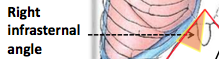
Right infrasternal angle |
pleura descending below the costal margins pleural site prone to injury infrasternal angle- site for pericardiocentesis (5th/6th left intercostal space) |
|
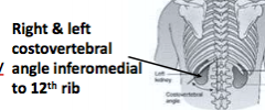
Costo-vertebral angle |
pleura exposed covering 12th rib during surgery, gets damaged, ex) kidney surgery |
|
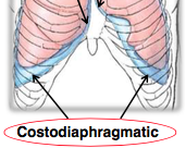
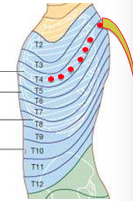
Pleural recess 2 |
Ideal site for thoracentesis/pleural tap only contains pleural fluids. Located superiorly to diaphragmatic dome. More pronounced from 8th to 10th ribs |
|
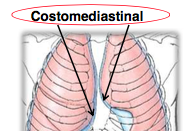
Pleural recess 1 (no clinical correlate; just know) |
Located posteriorly to sternum Left recess markedly larger than on right side because of cardiac notch |
|
|
Pleuritis |
inflammation of pleura irritation of parietal pleura innervated by intercostal nerves sharp chest pain, deep breath, cough auscultate and hear ROUGH, grating breathing sounds like fingers rubbing a hair strand |
|
|
Hydrothorax |
fluid in pleural cavity |
|
|
Hemothorax |
blood in pleural cavity |
|
|
Hemopneumothorax |
air and blood in pleural cavity |
|
|
Pyothorax |
pus in pleural cavity |
|
|
Chylothorax |
lymph in pleural cavity ~thoracic duct lymph |
|
|
Pleurodensis |
treatment for pneumothorax AND effusion closing of the pleural cavity |
|
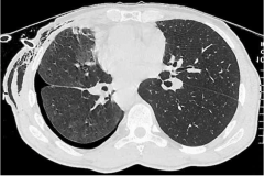
Pneumothorax |
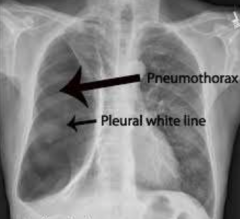
Air entry into pleural cavity 1. trauma (bullet wound, stab)- thoracic wall opening- tension type 2. bronchopulmonary fistula- a communication between lung &pleural cavity //congenital - open type
Treatment: pleurodensis |
|
|
Pleural Tap |
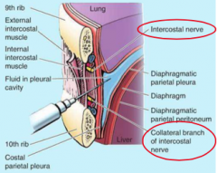
7th midclavicular, 9th midaxillary 10th paravetebral intercostal space wherever you feel comfortable counting ribs
Avoid diaphragm and point needle UPWARDS when taking pleura out of pleural cavity- costodiaphragmatic recess****
|
|
|
Chest Tube/Chest drain |
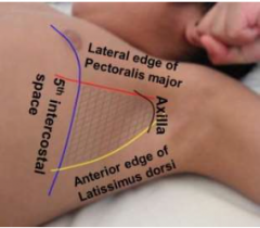
5th or 6th intercostal space at midaxillary line at nipple line "safe triangle" 1. Fluid removal: Tube directed inferiorly towards costodiaphragmatic recess 2. Air removal: Tube directed superiorly towards cervical pleura |
|
|
When do we need anesthesia to stop pain from occurring in thoracic wall? Where? Which nerves? |
When? Nerve block, Chest tube/drain, Pleural Tap (Thoracentestis) Where? Pleural tap (Thoracentesis) & Chest tube- skin, superficial fascia, 3 intercostal muscles, endothoracic fascia, and parietal pleura Nerve block: btwn internal and innermost ntercostal muscles Which nerves? intercostal nerves- sensitive to pain |
|
|
Extrapleural Intrathoracic Surgical Access |
Endothoracic fascia- Natural parietal cleavage plane to separate parietal pleura from inner thoracic wall |
|
|
Sibson's fascia |
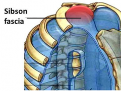
thickened endothoracic fascia protects cervical pleura/cupula that is prone to injuries above first rib to C7 |