Reading...
![]()
Play button
![]()
Play button
![]()
Use LEFT and RIGHT arrow keys to navigate between flashcards;
Use UP and DOWN arrow keys to flip the card;
H to show hint;
A reads text to speech;
91 Cards in this Set
- Front
- Back
|
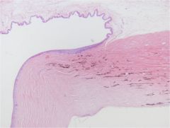
Ignore this slide b/c it is having issues!
|
|
|
|
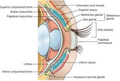
the opening of the eye
|
palpebral fissure
|
|
|
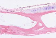
eyelid structure
|
Haired skin on the outside
Skeletal muscle - Orbicularis oculi Tarsal plate - Meibomian glands |
|
|
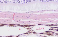
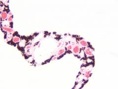
most common eyelid tumor in dogs
|
meibomian gland adenoma
|
|
|
meibomian glands
|
(tarsal gland)
sebaceous gland of the eyelid |
|
|
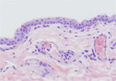
conjunctival epithelium
|
Stratified columnar or stratified squamous epithelium with goblet cells
- Goblet cells contribute to tear film - May be pigmented - Transitions to haired skin at the eyelid margin - Transitions to corneal epithelium at the limbus Palpebral: Lining or facing the eyelid Bulbar: Lining or facing the globe |
|
|
conjunctival epithelium
|
|
|
third eyelid
|
AKA: nictitans; nictitating membrane
Cartilage core Lined on both sides by conjunctival epithelium Palpebral (eyelid side) Bulbar (globe side) Contains lymphoid tissue Variably opaque - Species differences Associated lacrimal gland - AKA: gland of the 3rd eyelid - Mucous and serous components |
|
|
tear film
|
Meibomian glands (lipid component)
Lacrimal glands - Gland of the 3rd eyelid - Dorsolateral margins of the orbit - Compound tubuloalveolar structure |
|
|
orbit
|
Bony structure
Lacrimal glands - dorsolateral & orbital lacrimal glands Muscles Adipose tissue |
|
|
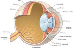
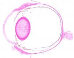
organization of the globe
(3 tunics) |
Fibrous tunic
- Cornea - Sclera Vascular tunic - Anterior uvea - Posterior uvea Nervous tunic - Retina |
|
|
fibrous tunic
|
cornea
sclera |
|
|
vascular tunic
|
anterior and posterior uvea
|
|
|
nervous tunic
|
retina
|
|
|
know the layout of the eye and the 3 tunics
|
|
|
|
_____ forms the outside of the globe
|
fibrous tunic
|
|
|
cornea
sclera pupil iris ciliary body zonular fibers (imagine them) lens anterior & posterior chambers iridocorneal angle vitreous chamber retina optic nerve ora ciliaris retina anterior and posterior uvea |
|
|
What is the junction of the sclera and cornea called?
|
limbus
|
|
|
cornea
|
Continuous with sclera
- Junction of sclera & cornea = limbus 3 main layers - Epithelium - Stroma - Endothelium Transparent |
|
|
Where is the highest concentration of nerves in the body?
|
corneal stroma
|
|
|
corneal stroma
|
Keratocytes (not keratinocytes)
Layers of collagen - White spaces (normal artifact) Avascular - Vessels form with disease Highest concentration of nerves in the body - Very sensitive - Can’t see with routine staining |
|
|
where are keratocytes found?
|
corneal stroma
|
|

|
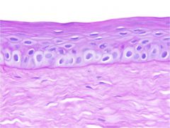
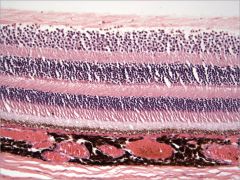
cornea
|
|
|
cornea
top (outside) epithelium stroma endothelium bottom (inside) |
|
|
corneal endothelium
simple squamous epithelium sits under basement membrane |
|
|
Corneal endothelium under descemet's membrane
|
|
|
corneal epidermalization
dried out cornea - rete pegs - keratin - inflammatory cells - blood vessels |
|
|
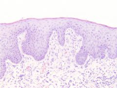
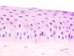
corneal epithelium
non-keratinizing stratified squamous |
|
|
corneal epithelium (PAS stain)
no keratin, pigment, adnexia bright pink line is basement membrane |
|
|
corneal nerves
|
|
|
corneal stroma
|
|
|
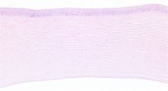
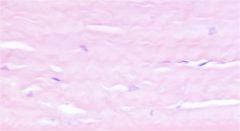
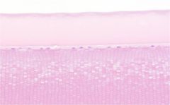
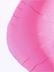
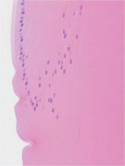
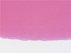
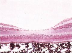
connective tissue 'capsule'
|
sclera
|
|
|
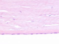
connective tissue of the sclera consists of
|
Nerves & blood vessels
birds - cartilage &/or bone |
|
|
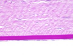
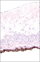
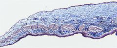
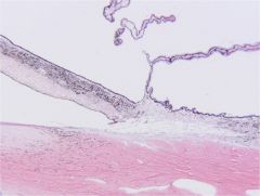
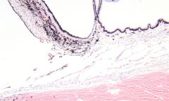
top left = conjunctival epithelium
bottom left = cornea right = sclera pigmented line in middle = limbus |
|
|
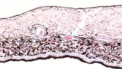
avian sclera
scleral ossicles (bone) pneumatic bone (air spaces) hyaline cartilage |
|
|
describe the pupil
|
The empty space bordered by the iris
Changes shape/size... dogs change size but are always round cats change shape and size horse has corpora nigracans which serves as a visor |
|
|
anterior uvea consists of ______
posterior uvea consists of ______ |
Anterior uvea
- Iris - Ciliary body Posterior uvea - Choroid - Nourishes retina |
|
|
How do you change the size/ shape of the pupil
|
muscles of the iris contract and relax
|
|
|
Iris
|
Fibroblasts
Collagen Melanocytes Blood vessels Lymphatics Nerves Muscles Epithelium 2 layers of pigmented epithelium on the posterior surface |
|
|
iris
notice artery, vein, nerve, & pigmented epithelium on posterior surface |
|
|
iris of a blue eye
|
|
|
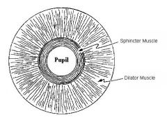
which direction does each iris muscle run?
|
|
|
|
iris muscles (trichrome stain)
sphincter muscle (thicker and higher up) dilator muscle (thinner at bottom) |
|
|
iris muscles (trichrome stain)
sphincter muscle (thicker and higher up) dilator muscle (thinner at bottom) |
|
|
ciliary body (anterior uvea)
notice ciliary processes & ciliary muscle sclera at bottom |
|
|
ciliary processes
pigmented (outer epitheliel layer) non-pigmented (inner epithelial layer) |
|
|
ciliary processes
|
Two layers of epithelium
- Non-pigmented - Pigmented (deeper layer) Continuous with the epithelium on the posterior surface of the iris Make the aqueous humor |
|
|
______ makes the aqueous humor
|
ciliary processes
|
|
|
what type of muscle is ciliary muscle?
|
smooth in mammals
skeletal in non-mammals |
|
|
ciliary process
inner epithelial layer up top smooth muscle throughout |
|
|
ciliary process in blue eyed dog
|
|
|
zonule fibers (pretend you see them)
Arise from Ciliary Body and attach to margins of the lens Contraction of ciliary body muscles adjust the shape of the lens for focusing light on different parts of the retina |
|
|
describe the lens
|
Held in place by zonule fibers
Avascular - no blood vessels, lymphatics, or nerves - Nourished by the aqueous humor anterior side has lens epithelial cells lens capsule surrounds lens lens fibers throughout lens bow (equator where epithelial cells move into the lens and then anterior) |
|
|
anterior lens capsule (gets thicker with age)
lens epithelial cells lens fibers (corn on the cob) |
|
|
lens bow
anterior (top) posterior (bottom) |
|
|
lens bow
|
|
|
What happens to the lens as you age?
|
Lens fibers are continually made and laid down on top of each other
Lens gets denser as you age |
|
|
posterior lens
no lens epithelial cells thin lens capsule |
|
|
|
posterior lens
no lens epithelial cells thin lens capsule |
|
|
know the structures of the eye
|

|
|
|
annular pad in birds
|
Lens fibers in the annular pad are arranged radially instead of concentrically
Allows for more accommodation (of the lens) = better vision |
|
|
describe the anterior chamber
|
Space behind the cornea and in front of the iris leaflets
Contains aqueous humor (made by ciliary processes) |
|
|
describe the posterior chamber
|
Space behind the iris leaflets and in front of the front half of the lens
Contains aqueous humor (made by ciliary processes) |
|
|
_____ is where aqueous humor flows out of the eye
|
iridocorneal angle
|
|
|
all aqueous humor leaving the eye eventually ends up draining to ______
|
the external jugular
|
|
|
iridocorneal angle
top left - iris bottom left - limbus bottom right - scleral plexus top right - ciliary body middle right - trabecular meshwork middle - small pectinate ligament |
|
|
iridocorneal angle (blue eye)
bottom - sclera top left - iris middle left - pectinate ligaments middle right - trabecular meshwork middle top - ciliary body |
|
|
normal aqueous outflow
|
Ciliary processes → posterior chamber → through pupil → anterior chamber → pectinate ligaments → trabecular meshwork → scleral plexus → venous system
|
|
|
glaucoma is a result of
|
an obstruction to aqueous outflow
|
|
|
Describe the vitreous chamber and it's contents
|
AKA: Posterior Compartment
- NOT posterior chamber Filled with vitreous humor - Gelatinous - Transparent - Helps hold the lens & retina in place |
|
|
describe the retina
what is it composed of |
Nervous tunic
Receives and transduces light and converts the information to nerve impulses which are sent to the brain via the optic nerve Composed of: - Photosensitive retina - Pigmented and non-pigmented retinal epithelium (RPE) |
|
|
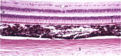
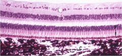
top (lighter purple) - retina
middle (dark) - choroid (posterior uvea) bottom (pink) - sclera |
|
|
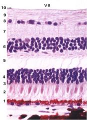
retina
3 nuclear layers - top: ganglion cell layer - middle: inner nuclear layer - bottom: outer nuclear layer |
|
|
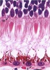
photoreceptor
M- outer limiting membrane notice cones and retinal pigmented epithelium (with melanin granules) |
|
|
describe retinal pigmented epithelium
|
Cuboidal epithelium
- With basement membrane Not always pigmented… Eats (prunes) and interacts with photoreceptor outer segments (rods & cones) |
|
|
if there are retinal blood vessels, where will they be found?
|
in the inner retina
|
|
|
retina
notice blood vessel in inner retina |
|
|
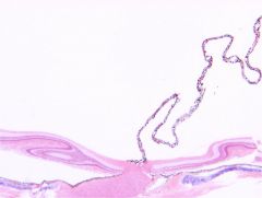
ora ciliaris retina
|
AKA: Ora serrata
Where the retina ends (nervous tunic) and the ciliary body begins... |
|
|
what is found between the retina and sclera?
What is its function? |
Choroid (posterior uvea)
- Nourishes retina |
|
|
The choroid consists primarily of _______
|
blood vessels and support tissue
pigmented in most animals |
|
|
tapetum lucidum
|
Layer of reflective tissue in the dorsal portion of the choroid of most domestic animals
Reflects the light that has just passed through the retina and restimulates the retinal photoreceptor cells Enhances night vision The RPE overlying the tapetum is NOT pigmented |
|
|
dog tapetum lucidum
Holangiotic vessels |
|
|
horse tapetum lucidum
Paurangiotic vessels |
|
|
tapetum lucidum
carnivore vs herbivore |
Carnivores: Brick-shaped cells containing reflecting crystals (tapetum cellulosum)
- Dog 15 layers - Cat 35 layers Herbivores: Regularly arranged collagen (tapetum fibrousum) |
|
|
tapetum fibrosum is found in
|
herbivores
|
|
|
Cat Tapetum
top = outer nuclear layer retina photoreceptors retinal pigmented epithelium (very thin) tapetum cellulosum (brick wall) choroid (lots of pigment and blood vessels) |
|
|
fovea and macula
|
Found in primates, birds, reptiles...and others...
NOT dogs, cats Paired structures... Fovea – depressed area in retina - Only photoreceptors and outer nuclear layer - Less refractory distortion Macula – surrounds fovea - All the cells that aren’t in the fovea - Thicker inner nuclear layer and ganglion cell layers - Macular degeneration... |
|
|
fovea (depressed area in middle)
macula (all the cells bunched up on the sides) get less visual distortion |
|
|
primate macula
|
|
|
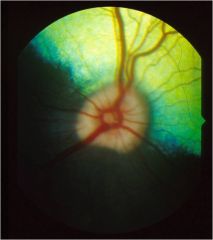
avian pecten
- nourishes the inner retina and inner eye bottom middle - optic nerve bottom right - sclera with cartilage above sclera - retina string of cells hanging off - pecten |
|
|
avian pecten
- nourishes the inner retina and inner eye |