Reading...
![]()
Play button
![]()
Play button
![]()
Use LEFT and RIGHT arrow keys to navigate between flashcards;
Use UP and DOWN arrow keys to flip the card;
H to show hint;
A reads text to speech;
79 Cards in this Set
- Front
- Back
|
What are the four main articulations in the tarsal (hock) joint?
|
Tarsocrural joint
Proximal Intertarsal joint Distal intertarsal joint Tarsometatarsal joint |
|
|
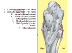
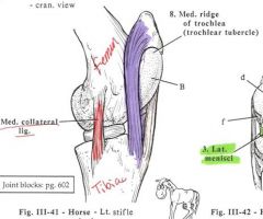
Which ridge of the trochlea of the femur is most important in patellar lock?
|

The larger medial ridge
|
|
|
Which synovial sac is most important and communicates with the proximal intertarsal sac?
|
tibiotarsal sac
|
|
|
ligament that stabilized the calcaneus when under force of common calcaneal tendon:
|

Long plantar lig.
|
|
|
What end of the common joint capsule is thin and often bulges out with synovial fluid accumulation (bog spavin)?
|
Proximal end
|
|
|
tendon of the cranial tibial mm
|
cunean tendon
|
|
|
Where should you inject the common joint capsule for the tarsocrural joint and proximal intertarsal joint?
|
Dorsal pouch
|
|
|
All tendons passing over the hock are enclosed in tendon sheaths except:
|
tendon of the super digital flexor
|
|
|
How many synovial sacs are there in the hock?
|
Four
|
|
|
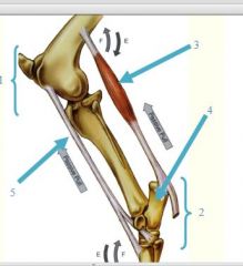
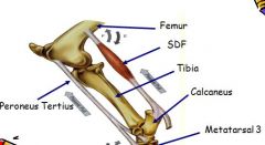
reciprocal apparatus ensures what?
|
simultaneous, reciprocal movement at both the stifle and hock joints, such that when the stifle joint flexes, the hock joint also flexes, the same with extension. The move in unison.
|
|
identify parts of stifle jt. shown
|
see picture
|
|
identify parts of stay aparatus
|
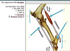
see picture
|
|
|
classify hip joint
|
HIP (COXAL) JOINT is Synovial, composite
ball and socket (spheroidal) joint |
|
|
ligament of the head of the femur - is very stout; extends from ___ to ____ ?
intra-articular portion enclosed in _____ ? |

extends from the acetabular fossa
to the fovea capitis of the femur); intra-articular portion enclosed in the synovial membrane. |
|
|
what muscle tendinous ?
|
SDF, DDF, and peroneus tertius
the tendinous interosseus arises proximal to fetlock ideal for weight bearing w/o much muscle activity, energy expended; support joints and prevent over extension gluteus profundus has numeous tendinous bands (unrelated) |
|
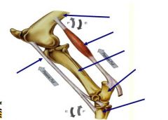
identify parts of reciprical ap. shown:
|
see picture
|
|
|
List ligaments of the hip joint.
|
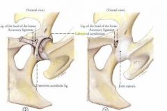
Lig. of head of the femur, the accessory lig., and transverse acetabular lig. and labrum acetabulum (acetabular lip) which deepens acet.cavity
|
|
|
When the hip joint is fully extended, there is a considerable amount of pull on the lateral crus of the superficial inguinal opening, predisposing horses to inguinal hernia, this connection to the medial thigh fascia by strands of connective tissue, present only in the horse. Is called:
|
femoral lamina
|
|
|
What ligament of the hip joint limits horse from kicking to the side?
|
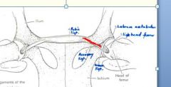
Accessory ligament
|
|
|
What are the movements of the hip joint?
|
Mainly extension and flexion
|
|
|
Where does the ligament of the head of femur attach in the acetabulum?
|

Acetabular fossa
|
|
|
What bones are involved in the hip joint?
|
Ilium
Pubis Ischium Head of Femur |
|
|
Accessory ligament extends from
|

the prepubic tendon, passing outwards, backwards and upwards, and through the acetabular notch to end in the fovea capitis, close to the ligament of the head of the femur
|
|
|
unlock stifle joints
|
Biceps femoris m.
|
|
|
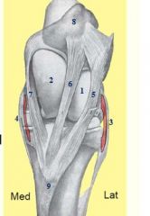
What muscles are part of the common calcaneal tendon?
|
Biceps femoris muscle
Gastrocnemius muscle Superficial digital flexor muscle Gracilis muscle Semitendinosus muscle Soleus muscle |
|
|
What can inflammation in the calcaneal bursae cause?
|
Capped hock
|
|
|
Synovial bursa present in between attachments of accessory gluteal muscle?
|
Trochanteric Bursa:
Synovial bursa present in between cranial part of greater trochanter and aponeurotic attachment of *accessory gluteal muscle* |
|
|
What is the normal angle of flexion in the hip joint?
|
110 - 115 degrees, absorbs concussion
|
|
|
What is the largest single joint of the body?
|
Stifle joint
|
|
|
The stifle joint is a compound joint, what are the two joints and what kind of joints are they?
|
Femoropatellar joint (simple gliding)
Femorotibial joint (simple condylar) |
|
|
What are the bones associated with the stifle joint?
|
Distal femur
Proximal tibia Patella |
|
|
Which nerve supplies the quadriceps mm?
Function? |
Femoral n
Extension if stifle and support, pulling up patella to lock it (wt bearing) flexes hip (rectus femoris) |
|
|
What are the three joint capsules of the stifle joint?
|

Femoropatellar
Medial femorotibial Lateral femorotibial |
|
|
What ridge is largest in the trochlea of the distal femur?
|
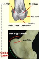
Medial ridge
|
|
|
What are the two types of surfaces on the trochlea of the distal femur?
|
Resting surface
Gliding surface |
|
|
What are the three synovial sacs in the stifle joint?
|
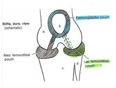
Femoropatellar sac
Lateral femorotibial sac Medial femorotibial sac |
|
|
Which two synovial sacs in the stifle joint communicate 100% of the time?
|

Femoropatellar sac
Medial femorotibial sac |
|
|
Which two synovial sacs in the stifle joint communicate in only 25% of horses? (not very much)
|
Femoropatellar sac
Lateral femorotibial sac |
|
|
Which two synovial sacs in the stifle joint never communicate?
|

Lateral femorotibial sac
Medial femorotibial sac |
|
|
What is the largest sesamoid bone of the body?
|
Patella
|
|
|
Where on the patella is the parapatellar fibrocartilage found?
|
Medial angle
|
|
|
What two services does the patella provide the quadriceps femoris muscle?
|
Provides a large gliding surface over the trochlea of the femur
Protects the tendon of insertion from excessive friction |
|
|
functional digit of the horse
|
digit 3
|
|
|
What is the purpose of the medial and lateral menisci in the femorotibial joint?
|
minimizes incongruence b/t the two bones
|
|
|
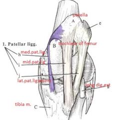
What are the three tendons of the patella?
|

Medial patellar ligament
Middle patellar ligament Lateral patellar ligament |
|
|
How many collateral ligaments are in the stifle joint of the horse?
|
2 (medial and lateral)
|
|
|
How many cruciate ligaments are there and what is their function?
|
Cranial and Caudal
Stabilize the stifle joint |
|
|
How many meniscal ligaments are there and what is their funciton?
|
4 ligaments
Attache cranial and caudal horns of the menisci to the cranial and caudal intercondylar areas |
|
|
What ligaments limit hyper-extension and hyperflexion?
|
Hyper-extension - cruciates (especially cranial)
Hyper-flexion - cruciates |
|
|
How many femoropatellar ligaments are there and what is their function?
|

Medial and lateral
Attach parapatellar fibrocartilage to the femoral epicondyles |
|
|
Is the stifle joint normally partially flexed?
|
Yes, concussion absorbing
|
|
|
When does the patellar lock occur?
|
When the horse bears body weight only on one limb
|
|
|
What is the angle of the limb when the patellar lock occurs? Normally?
|
Patellar lock - 145 -150 degrees
Normal - 135 degrees |
|
|
What muscle action unlocks the patella?
|
Biceps femoris muscle (cranial division)
|
|
|
What ligament can be cut in the stifle joint and not allow the animal to lock the limb?
|
Medial patellar ligament
|
|
|
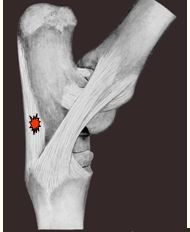
Where can the lateral femorotibial joint cavity be injected?
|
B/t long digital extensor tendon and lateral collateral ligament
|
|
|
Where can the medial femorotibial joint cavity be injected?
|

B/t medial patellar ligament and medial collateral ligament
|
|
|
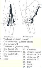
How many bones are in the tarsal (hock) joint?
|
6 bones
|
|
|
Where can the femoropatellar joint cavity be injected?
|

Caudal to the lateral patellar ligament
|
|
|
What bones are located in the proximal row of the hock joint?
|
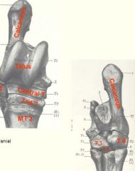
Talus
Calcaneus |
|
|
What bones are located in the middle and distal rows of the hock joint?
|

T1 and T2
T3 and T4 |
|
|
What helps the stifle avoid the caudal thoracic wall during flexion?
|
Obliquity of the trochlea
|
|
|
Where is the medial collateral ligament of the hock?
|
From medial malleolus to the talus and proximal ends of the 2nd and 3rd metatarsals
|
|
|
Where is the lateral collateral ligament of the hock?
|
From lateral malleolus to the calcaneus, fourth tarsal, and 3rd and 4th metatarsal
|
|
|
What ligament stabilizes the calcaneus when under force of common calcaneal tendon?
|
Long plantar ligament
|
|
|
What ligament arises on the distal tuberosity on the medial part of the talus and inserts on the 2nd and 3rd metatarsal?
|
Dorsal ligament
|
|
|
What ligament is a distal extension of the plantar part of the joint capsule and joins the tendon of the deep digital flexor muscle?
|
Accessory ligament
|
|
|
Where does the dorsal limb of the cunean tendon insert on?
|

Metatarsal 3
|
|
|
Where does the medial limb of the cunean tendon insert on?
|
Tarsals 1 and 2
|
|
|
What bursa lies under the cunean tendon and over the medial collateral ligament of the hock?
|
Subtendinous or cunean bursa
|
|
|
The condition known as curb, is when this tendon is swollen due to strain and causes convexity of plantar surface?
|

Long plantar ligament
|
|
|
What tendons are located in the tarsal synovial sheath?
|
Tendon of lateral digital flexor m.
Two of the three heads of the deep digital flexor m. |
|
|
Is the hock joint usually partially flexed?
|
Yes, concussion-absorption
|
|
|
What is the hardest worked joint in the equine body?
|
Hock joint
|
|
|
What does strain and stress on the hock joint cause?
|
Spavin
|
|
|
What is the cranial portion of the reciprocal apparatus in the hindlimb?
|
Peroneus (fibularis) tertius m.
|
|
|
What is the caudal portion of the reciprocal apparatus in the hindlimb?
|
Superficial digital flexor tendon (almost entirely tendinous)
|
|
|
If the peroneus tertius muscle ruptures what happens to the hock?
|
Abnormal, jerky flexing of the hock
|
|
|
What happens if both the peroneus tertius muscle and digital flexor tendons is ruptured?
|
hock drops to the ground
|