Reading...
![]()
Play button
![]()
Play button
![]()
Use LEFT and RIGHT arrow keys to navigate between flashcards;
Use UP and DOWN arrow keys to flip the card;
H to show hint;
A reads text to speech;
56 Cards in this Set
- Front
- Back
|
How many segments are in the spinal cord?
|
31: 8C+12T+5L+5S+1Co
|
|
|
The spinal cord has two enlargements located where?
|
cervical and lumbar regions. These are big thanks to the upper and lower limbs.
|
|
|
In the adult the spinal cord ends at ...
|
L1 - L2
|
|
|
What ends at S2, the level of which is marked by a line through the posterior superior iliac spines (PSISs).
|
Dural sac or thecal sac
|
|
|
The spinal cord is attached distally to the coccyx by what structure?
|
filum terminale.
Internal filum is formed by pia mater. External filum has a covering of dura. |
|
|
What causes muscle weakness in multiple muscles rather than paralysis of those muscles?
|
radiculopathy, (single nerve damage) Because a myotome contributes to more than one muscle
|
|
|
Peripheral nerve injuries affect more than one dermatome and are also known as what?
|
neuropathy
|
|
|
Peripheral nerve injuries affect adjacent dermatomes and cause ____ of the innervated muscles
|
paralysis
|
|
|
why is white matter white?
|
because of the myelinization by oligodendroglial cells.
It contains tracts, which are bundles of myelinated axons. |
|
|
What is contained in the gray matter?
|
neuronal cell bodies and dendrites.
|
|
|
where do lower motor neurons reside?
|
anterior horn of spinal cord.
Their axons innervate skeletal muscle Their axons form the motor portions of peripheral nerves They are called “the final common pathway” because they receive input from higher brain areas such as the cerebral cortex |
|
|
Where do upper motor neurons reside?
|
Upper motor neurons are in higher centers such as the motor cortex
Their axons excite or inhibit lower motor neurons |
|

What is this picture representing?
|
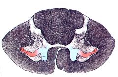
There is a topographic representation of the limb (somatotopic).
Generally, the lower motor neurons that innervate axial muscles are more medial. The lower motor neurons innervating the proximal limb muscles are lateral to those and the lower motor neurons innervating the more distal limb muscles are most lateral. Lower motor neurons innervating flexors are more dorsal (posterior) and those innervating extensors are more ventral (anterior). |
|
|
Stroke the skin of the abdomen, underlying muscles will contract.
What spinal segments are involved? |
This is a superficial reflex.
Spinal segments Upper abdomen: T7-T9 Lower abdomen: T10-T11 |
|
|
Cremasteric reflex involves stroking the skin of the medial thigh. Cremasteric muscle contracts elevating testis.
What spinal segments does this test? |
Tests T12, L1 and L2
|
|
|
Plantar reflex involves stroking bottom of foot from heel to toe. Provokes plantar flexion of toes.
What spinal segments does this test? |
Tests L4, L5, S1 and S2
|
|
|
“Anal Wink” involves stimulus of perianal region. Anal sphincter contracts.
What spinal segments does this test? |
Tests S2-S4
|
|
|
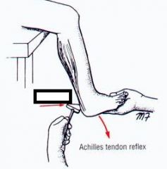
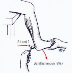
The following reflex tests what spinal segments:
Ankle(Achilles) reflex |
S1 and S2
|
|
|
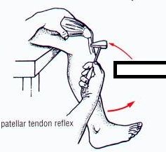
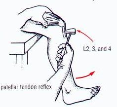
The following reflex tests what spinal segments:
Knee (patellar) |
L2, L3 and L4
|
|
|
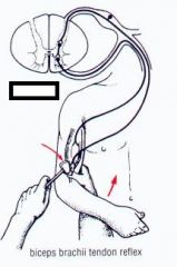
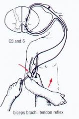
The following reflex tests what spinal segments:
Biceps reflex |
C5 and C6
|
|
|
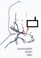
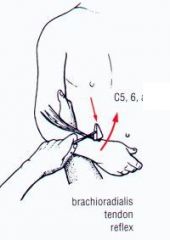
The following reflex tests what spinal segments:
Brachioradialis reflex |
C5 and C6
|
|
|
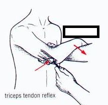
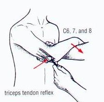
The following reflex tests what spinal segments:
Triceps reflex |
C6, C7 and C8
|
|
|
|
|
|
|
|
|
|
|
|
|
|
|
|
|
|
What are the basic steps involved in the deep tendon reflexes?
|
Muscle spindles detect muscle length and stretch.
When a muscle is stretch by tapping its tendon with a reflex hammer, that information is carried to the spinal cord by a 1a afferent axon. The 1a fiber is a proprioceptive afferent, i.e. carries information about deep somatic structures. The 1a fibers synapse directly on alpha motor neurons that innervate the muscle (monosynaptic). The alpha motor neurons fires and the muscle contracts. Alpha motor neurons innervating the antagonist muscle are inhibited. |
|
|
Clinically, the most important descending tract, The lateral corticospinal tract, starts where and ends where?
|
Arises from upper motor neurons in the motor cortex
(Motor cortex is somatotopically organized) Axons descend and cross in the pyramidal decussation in the caudal medulla Innervates lower motor neurons which innervate limb muscles |
|
|
What are two major sensory pathways for the somatosensory system?
|
Dorsal columns (lemniscal system)
Anterolateral (spinothalamic) system |
|
|
Of the Two major sensory pathways, what sensory info does the Dorsal columns (lemniscal system) pathway send and where does it cross?
|
discriminative touch, proprioception, and vibratory sense
Crosses in the medulla |
|
|
Of the Two major sensory pathways, what sensory info does the anterolateral (spinothalamic) pathway send and where does it cross?
|
crude touch, pain and temperature
Crosses in the spinal cord |
|
|
Injury of corticospinal system (pyramidal tract) anywhere above the pyramidal decussation causes ___ paralysis paresis of the limbs.
|
contralateral
|
|
|
upper motor neuron Injury below the pyramidal decussation will cause ____ paralysis or paresis below the lesion.
|
ipsilateral
|
|
|
Injury to the spinal cord will cause loss of pain and temperature sense on the ____ side below the lesion.
|
contralateral
|
|
|
An injury to the spinal cord will cause _____ loss of fine (discriminative) touch, proprioception and vibration below the lesion.
|
ipsilateral
|
|
|
A lesion that involves ½ of the spinal cord at T6 vertebral level will cause:
Paralysis of the _____ lower body. _____ loss of fine touch (2 point discrimination), proprioception and vibration sense below the lesion (lower ½ of body) ______ loss of pain and temperature below the lesion (opposite side of lower body) |
ipsilateral
Ipsilateral Contralateral |
|
|
What are three layers of the meninges?
|
dura mater, arachnoid, pia mater
|
|
|
What is Separated from the periosteum of the vertebrae by the epidural space?
|
dura mater
|
|
|
What is Separated from the dura by a potential space, the subdural space?
|
arachnoid
|
|
|
what is Tightly adherent to cord, Forms denticulate ligaments, Forms internal filum terminale?
|
pia mater
|
|
|
Dura mater is derived from what?
|
mesenchyme, hence the alternate name, pachymeninx
|
|
|
the dura mater is made up of:
|
Dense CT
Longitudinal collagen fibers with circular elastic fibers |
|
|
the Leptomeninx consists of what two layers?
|
the arachnoid and pia mater
|
|
|
the Leptomeninx is derived from embryonically?
|
neural crest
|
|
|
True or False: Arachnoid mater and pia mater both contain blood vessels
|
True
|
|
|
where does the thecal sac end? and what is it enclosed by?
|
the thecal sac is also known as the dural sac as it is made of dura mater. it ends at s2.
|
|
|
Below the S2 level, after the thecal/dural sac has closed, the dura continues as the ____ which then ends at the coccyx.
|
filum terminale
|
|
|
The subdural space is a potential space, which means:
|
One cannot enter the subdural space with a needle in the living patient.
|
|
|
What is the most definitive index of meningitis and how do you get this?
|
The presence of Polymorphonuclear leukocytes, which can be gained by a sample of CSF.
|
|
|
Where is the needle inserted for a lumbar puncture or spinal tap?
|
Needle inserted through the ligamentum flavum between L3 and L4 or between L4 and L5 (Caudal to the spinal cord)
ALSO Roots of cauda equina are not damages they are pushed aside within the CSF |
|
|
What is an epidural?
|
An Anesthetic infused around outside of dural sac which Allows selective block of roots without affecting cord
|
|
|
What is a normal anion gap?
|
4-12
|
|
|
Lower 2/3 of spinal cord dependent on a medullary segmental artery that is usually larger than the others, which typically (65% of people) arises on the left at T12 or L1. What is this called?
|
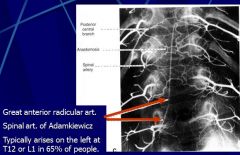
, the arteria radicularis magna or great radicular artery of Adamkiewicz
|
|
|
Explain the clinical significance and characteristics of Metastases via the Epidural (Batson’s) Plexus?
|
The epidural plexus that drains the spinal cord and meninges has no valves
Increased intra-abdominal pressure can cause a reflux of blood into the plexus and thereby carry metastatic cells, such as prostate CA and infections, such as pelvic infections, along the spinal column to more cranial levels. |
|
|
Case Presentation: A 28-year-old man was drinking around the swimming pool with friends and, on a dare, dove into the pool and struck his head on the bottom. When pulled out by friends, he couldn’t move his left upper or lower limb and, when asked, he could not wiggle the toes on his left foot or move the fingers of his left hand. He was rushed to the Emergency Department where an examination revealed left sided paralysis below the neck, left sided loss of fine touch, proprioception and vibratory sense, and right-sided loss of pain and temperature, all below the neck.
Where is his injury? Where do the affected tracts cross? |
Left cervical spinal cord
Corticospinal and dorsal column system –caudal medulla Anterolateral system – in the spinal cord |