Reading...
![]()
Play button
![]()
Play button
![]()
Use LEFT and RIGHT arrow keys to navigate between flashcards;
Use UP and DOWN arrow keys to flip the card;
H to show hint;
A reads text to speech;
10 Cards in this Set
- Front
- Back
- 3rd side (hint)
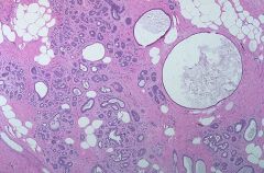
Diagnosis?
|
Fibrocystic Change of the Breast:
Note the combination of mutiple benign changes-fibrosis, microcysts, adenosis |
|
|
Diagnosis?
|
Cyst with apocrine change (Apocrine cyst)
How likely is this cyst to recur when compared to a simple cyst with cuboidal epithelium |
More likely to recur.
|
|
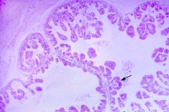
Diagnosis (2 synonomous names)
|
Blunt duct adenosis or Columnar cell change
Name the histologic criteria: |
luminal epith replaced by columnar cells with snouts. The ducts take on a more complicated pattern with branching, etc.
This is a benign entity |
|
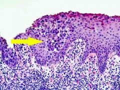
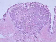
Diagnosis, please.
|
Paget's disease of the nipple
What is the most common underlying malignancy in an affected breast? |
Ductal carcinoma (sometimes DCIS!)
|
|
|
What are Toker cells?
|
Clear cells that can appear in the nipple and may be mistaken for Paget cells.
|
|
|
|
What are helpful stains in differentiating Paget cells from Bowen disease and Melanoma?
|
Bowen disease- Keratinocyte CK+, S100-, EMA-, Her2Neu-
Melanoma: melanin+, s100+, CK- Paget: Melanin +/-, CEA +, keratinocyte CK -, S100 +/-, H2Neu + |
|
|
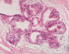
Identify:
|
Nipple Adenoma
Note the abrupt transition to the glandular tissue, distinct layers of myoep and epith tissue. |
|
|
|
A benign nipple lesion characterized by comma-shaped tubules
|
Syringomatous adenoma of the nipple
|
|
|
Identify:
|
Apocrine metaplasia
|
|
|
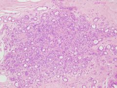
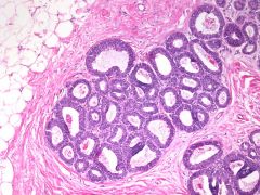
Identify:
|
Sclerosing Adenosis
Note the compression of the glands by the stroma but overall retention of the lobular structure and double-layered glands. What IHC can help you distinguish this from malignancy? |
Stains for the myoepithelial layer:
HMWK (CK5/6), SMA, Calponin, smooth muscle myosin heavy chain, p63 |