Reading...
![]()
Play button
![]()
Play button
![]()
Use LEFT and RIGHT arrow keys to navigate between flashcards;
Use UP and DOWN arrow keys to flip the card;
H to show hint;
A reads text to speech;
224 Cards in this Set
- Front
- Back
|
What four things is the nervous system composed of? |
brain, spinal cord, nerves, ganglia
|
|
|
What are the 3 general functions of the nervous system?
|
Collect information
Process and evaluate information Initiate response to information |
|
|
Specialized nervous system cells
|
neurons
|
|
|
_____ are specialized nervous system structures that monitor changes in both the internal and external environment called stimuli
|
Receptors
|
|
|
Changes to the internal and external environment is called _____
|
stimuli
|
|
|
_____ include all three types of muscle tissue and glands.
|
Effectors
|
|
|
What are the different types of effectors controlled by the nervous system?
|
All three types of muscle tissue and glands
|
|
|
What are the two anatomic divisions of the nervous system?
|
central nervous system (CNS)
peripheral nervous system (PNS) |
|
|
____ includes the brain and spinal cord
|
CNS
|
|
|
_____ includes nerves and ganglia
|
PNS
|
|
|
_____ are bundles of neuron processes (axons)
|
nerves
|
|
|
____ are clusters of neuron cell bodies located along nerves
|
ganglia
|
|
|
What are the two functional divisions of the nervous system?
|
sensory and motor
|
|
|
_____ is responsible for receiving sensor information FROM receptors that detect stimuli and transmitting this information TO the CNS.
|
sensory nervous system
|
|
|
How is the sensory nervous system subdivided?
|
somatic (conscious)
visceral (automatic) |
|
|
Receptors of the somatic sensory nervous system include:
|
eyes, nose, tongue, ears, skin, proprioreceptors
|
|
|
Receptors in muscle and joints that detect body position
|
proprioreceptors
|
|
|
_____ sensory components detect stimuli that we don't consciously perceive
|
visceral sensory components
|
|
|
Receptors of the visceral sensory nervous system include:
|
structures located within blood vessels and internal organs, e.g., heart, stomach, kidneys
|
|
|
What is another name for the motor nervous system?
|
efferent nervous system
|
|
|
The _____ is responsible for initiating and transmitting motor output FROM the CNS TO effectors
|
motor nervous system
|
|
|
This system controls muscle tissue and glands
|
motor nervous system
|
|
|
Is the motor nervous system subdivided into somatic and visceral parts
|
yes
|
|
|
The _____ component initiates and transmits motor output from the CNS to voluntary skeletal muscles
|
somatic motor
|
|
|
what is the other name for autonomic motor nervous system?
|
visceral motor
|
|
|
the visceral motor component of the motor nervous system innervates and regulates:
|
cardiac muscle, smooth muscle, and glands
|
|
|
What are the two subdivisions of the autonomic nervous system?
|
sympathetic; parasympathetic
|
|
|
_____ is the primary tissue of the nervous system.
|
Nervous tissue
|
|
|
nervous tissue is composed of two distinct cell types:
|
neurons and glial cells
|
|
|
_____ are excitable cells that initiate and transmit electrical signals.
|
neurons
|
|
|
_____ cells are nonexcitable cells that primarily support and protect neurons
|
glial cells
|
|
|
Neurons have several special characteristics including:
|
excitability
conductivity secretion extreme longevity amitotic |
|
|
This is a responsiveness to stimulation
|
excitability
|
|
|
The type of stimulation that a neuron responds to is dependent upon ______
|
its location
|
|
|
Most neurons usually respond only to binding of secreted molecules called _____
|
neurotransmitters
|
|
|
neurotransmitters are released from
|
other neurons
|
|
|
These are electrical changes that are quickly propagated along the plasma membrane of neurons following stimulation
|
conductivity
|
|
|
Neurons release neurotransmitters in response to _____
|
conductive activity
|
|
|
How many types of neurotransmitters does a typical neuron release?
|
only one type
|
|
|
a neurotransmitter may have either ___ or ___ effect on its target organ
|
excitatory or inhibitory
|
|
|
T/F
most neurons formed during fetal development are still functional in very elderly individuals |
True
|
|
|
When is mitotic development lost in most neurons?
|
during fetal development of neurons
|
|
|
What are the exceptions to loss of mitotic development of neurons during fetal development?
|
olfactory epithelium of the nose and certain areas of the brain
|
|
|
This region of the brain is involved in memory processing:
|
hippocampus
|
|
|
This region of the brain contains a population of neural stem cells
|
hippocampus
|
|
|
What are the basic structural features of a neuron?
|
cell body, dendrites, an axon
|
|
|
What is another name for a cell body?
|
soma
|
|
|
What is another name for a soma?
|
cell body
|
|
|
a cell body is enclosed by a _____
|
plasma membrane
|
|
|
a cell body has _____ surrounding a nucleus
|
cytoplasm
|
|
|
_____ serve as the neuron's control center
|
cell bodies
|
|
|
_____ conduct electrical signals to the axon
|
cell bodies
|
|
|
electrical signals can either be initiated _____ or _____
|
within the cell body;
received from the dendrites |
|
|
What is the cytoplasm within a neuron cell body called?
|
perikaryon
|
|
|
Ribosomes are formed in ___ in a neuron
|
nucleolus
|
|
|
Where is the nucleolus in a neuron?
|
The nucleus
|
|
|
_____ occurs at approx 400 millimeters per day and involves movement along microtubules.
|
Fast axonal transport
|
|
|
_____ occurs at apprix .1 - 3 millimeters per day and results from the flow of the axoplasm, also called axoplasmic flow.
|
Slow axonal transport
|
|
|
______ are the most common type of neuron
|
multipolar neurons
|
|
|
These neurons have many dendrites and a single axon that extends from the cell body.
|
Multipolar neurons
|
|
|
_____ have two processes that extend from the cell body - one dendrite and one axon.
|
bipolar neurons
|
|
|
The location of these neurons is relatively limited in humans eg retina of the eye and olfactory mucosa in the nasal cavity
|
bipolar neurons
|
|
|
In what two locations can bipolar neurons be found in humans?
|
retina of the eye, olfactory mucosa in the nasal cavity
|
|
|
What are the three categories of functional classification of neurons?
|
Sensory neurons
Motor neurons Interneurons |
|
|
Are most sensory neurons unipolar, bipolar, multipolar, or anaxonic? What are the exceptions?
|
unipolar
retina and nasal mucosa are bipolar |
|
|
All motor neurons are unipolar, bipolar, multipolar, or anaxonic?
|
multipolar
|
|
|
Where do the cell bodies fo the motor neurons lie?
|
In the CNS
|
|
|
What is another name for sensory neurons?
|
afferent neurons
|
|
|
What is another name for afferent neurons?
|
sensory neurons
|
|
|
What is another name for motor neurons?
|
efferent neurons
|
|
|
What is another name for efferent neurons?
|
motor neurons
|
|
|
what is another name for interneurons?
|
association neurons
|
|
|
what is another name for association neurons
|
interneurons
|
|
|
Interneurons lie entirely within the ___
|
CNS
|
|
|
_____ facilitate communication between sensory and motor neurons
|
interneurons
|
|
|
_____ outnumber all other neurons
|
interneurons
|
|
|
it is estimated that ____% of our neurons are _____
|
interneurons
|
|
|
Interneurons are generally multipolar, unipolar, bipolar, anaxonic
|
multipolar
|
|
|
A _____ is a cable-like bundle of parallel axons that are components of the PNS
|
nerve
|
|
|
A nerve is a cable-like bindle of parallel axons that are components of the _____
|
PNS
|
|
|
A thick layer of dense irregular connective tissue that encloses the entire nerve and provides both support and protection.
|
Epineurium
|
|
|
What is the epineurium?
|
A thick layer of dense irregular connective tissue that encloses the entire nerve and provides both support and protection
|
|
|
a layer of dense irregular connective tissue that wraps fascicles, which are bundles of axons. This layer supports blood vessels
|
perineurium
|
|
|
What is the perineurium?
|
a layer of dense irregular connective tissue that wraps fascicles, which are bundles of axons. This layer supports blood vessels.
|
|
|
This layer of connective tissue wrappings supports blood vessels
|
perineurium
|
|
|
An individual axon in a myelinated neuron is surrounded by _____, then wrapped in the _____
|
neurolemmocytes; endoneurium
|
|
|
What kind of connective tissue separates and insulates each axon as part of the endoneurium?
|
areolar
|
|
|
What separates and electrically insulates each axon?
|
endoneurium (areolar connective tissue)
|
|
|
Within which connective tissue layer lie capillaries that supply each axon?
|
endoneurium
|
|
|
_____ nerves extend from the brain
|
cranial nerves
|
|
|
_____ nerves extend from the spinal cord
|
spinal nerves
|
|
|
What are the two structural classifications of nerves?
|
cranial nerves and spinal nerves
|
|
|
What are the three functional classifications of nerves?
|
sensory nerves, motor nerves, mixed nerves
|
|
|
Sensory nerves relay information (to/from) the CNS
|
to
|
|
|
Motor nerves relay information (to/from) the CNS
|
from
|
|
|
What are the three connective tissue wrappings in a nerve, and what specific structure does each ensheathe?
|
Epineurium; entire nerve
perineurium; fascicles or bundles of axons endoneurium; each axon |
|
|
The specific location where a neuron is functionally connected to either another neuron or an effector
|
synapse
|
|
|
What is a synapse?
|
The specific location where a neuron is functionally connected to either another neuron or an effector
|
|
|
What are the two types of synapses in the human body?
|
chemical; electrical
|
|
|
Most synapses within the nervous system are _____ synapses (chemical, electrical)
|
chemical
|
|
|
A _____ synapse is composed of a presynaptic neuron, which is a signal producer, and a postsynaptic neuron, which is the signal receiver or target
|
chemical synapse
|
|
|
A chemical synapse is composed of a ______ which is a signal producer and a _____ which is the signal receiver
|
presynaptic neuron; postsynaptic neuron
|
|
|
in regards to a chemical synapse, which is the signal producer and which is the signal receiver or target?
|
presynaptic is the producer
postsynaptic is the receiver or target |
|
|
Transmission between a pre and post synaptic neuron occurs when _____ molecules stored in synaptic vesicles are released from the synaptic knob of a presynaptic neuron into the synaptic cleft.
|
neurotransmitter molecules
|
|
|
There is a ______ associated with neurotransmitter release at chemical synapses.
|
synaptic delay
|
|
|
What causes the synaptic delay?
|
release of neurotransmitter from presynaptic cell, the diffusion across the synaptic cleft, and the binding to receptors in the postsynaptic plasma membrane.
|
|
|
An _____ is composed of a presynaptic and postsynaptic neuron fused together
|
electrical synapse
|
|
|
_____ are present in the plasma membranes of both neurons and facilitate the flow of ions between cells in electrical synapses
|
gap junctions
|
|
|
Is there synaptic delay with electrical synapses?
|
no
|
|
|
Which has synaptic delay? Chemical synapses or electrical synapses?
|
chemical synapses
|
|
|
Where are electrical synapses in the body?
|
limited regions of the brain and the eyes
|
|
|
what kind of synapses are in limited regions of the brain and the eyes?
|
electrical synapses
|
|
|
Are glial cells excitable?
|
No
|
|
|
What is the primary function of glial cells?
|
to support and protect the neurons
|
|
|
What is another name for glial cells?
|
neuroglia
|
|
|
Are glial cells found in both the CNS and PNS?
|
yes
|
|
|
what is another name for neuroglia
|
glial cells
|
|
|
Are glial cells larger or smaller than neurons?
|
smaller
|
|
|
Are glial cells capable of mitosis?
|
Yes
|
|
|
Do glial cells transmit nerve signals?
|
no
|
|
|
The _____ cells cooperate to physically protect and help nourish neurons as well as provide an organized, supporting scaffolding for all the nervous tissue.
|
glial
|
|
|
During development, _____ cells form the framework that guides young, migrating neurons to their final destinations.
|
glial cells
|
|
|
Do glial cells outnumber neurons or vice versa
|
glial cells outnumber neurons
|
|
|
_____ account for roughly half of the volume of the nervous system
|
glial cells
|
|
|
Large cel with numerous cell processes; in contact with neurons and capillaries; most common type of glial cell
|
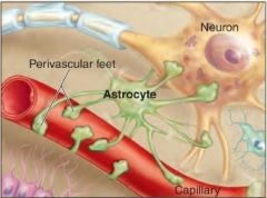
Astrocyte
|
|
|
Helps form the blood-brain barrierRegulates tissue fluid compositionProvides structural support and organization to the CNSAssists with neuronal developmentReplicates to occupy space of dying neurons
|

Astrocyte
|
|
|
Simple cuboidal or columnar epithelial cells that line cavities in brain and spinal cord; cilia on apical surface
|
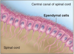
Ependymal cell
|
|
|
Lines ventricles of brain and central canal of spinal cordAssists in production and circulation of cerebrospinal fluid (CSF)
|

Ependymal cell
|
|
|
Small cell with slender branches from cell body; least common type of glial cell
|

Microglial cell
|
|
|
Defends against infectious agents and engulfs debris from dead or dying neurons
|
Microglial cell
|
|
|
Rounded, bulbous cell with slender cytoplasmic extensions; extensions wrap around CNS axons
|

Oligodendrocyte
|
|
|
Myelinates and insulates CNS axonsAllows faster action potential conduction along axons in the CNS
|

Oligodendrocyte
|
|
|
Flattened cell; groups of these cells cluster around neuronal cell bodies in a ganglion
|

Satellite cell
|
|
|
Protects and regulates nutrient and waste exchange for cell bodies in ganglia
|

Satellite cell
|
|
|
flattened cell wrapped around a portion of an axon in the PNS
|

Neurolemmocyte
|
|
|
Myelinates and insulates PNS axons and allows for faster action potential conduction along an axon in the PNS
|

Neurolemmocyte
|
|
|
What are the four types of glial cells found in the CNS?
|
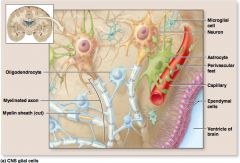
astrocytes, ependymal cells, microglia, and oligodendrocytes
|
|
|
astrocytes, ependymal cells, microglia, and oligodendrocytes are:
|

The four types of glial cells found in the CNS
|
|
|
These glial cells of the CNS have a star-like shape
|
astrocytes
|
|
|
Astrocyte glial cells of the CNS have projections that touch both _____ and _____
|
capillary walls, neurons
|
|
|
_____ are the most abundant glial cell in the CNS and constitute over 90% of the nervous tissue in some areas of the brain
|
Astrocytes
|
|
|
Astrocytes nurture, protect, support, and guide neurons in these 5 ways:
|
help form the blood - brain barrier
regulate tissue fluid composition form a structural network assist neuronal development occupy the space of dying neurons |
|
|
The ends of astrocyte processes are called _____
|
perivascular feet
|
|
|
What are perivascular feet?
|
The ends of astrocyte processes
|
|
|
The _____ and the _____ together contribute to a blood brain barrier (BBB)
|
perivascular feet; capillaries
|
|
|
The _____ strictly controls movement of tubstances from exiting the blood and entering the nervous tissue in the brain
|
Blood-brain barrier (BBB)
|
|
|
How do astrocytes help regulate the chemical composition of the interstitial fluid within the brain ?
|
controlling movement of ions and molecules between the blood and the interstitial fluid
|
|
|
The cytoskeleton in _____ strengthens and organizes nervous tissue in the CNS by forming a framework to support neurons
|
astrocytes
|
|
|
_____ help direct the development of neurons in the fetal brain by secreting chemicals that regulate the formation of connections between neurons
|
astrocytes
|
|
|
When neurons are damaged and die, the space they formerly occupied is often filled by cells produced by ______ _______, a process termed astrocytosis.
|
astrocyte division
|
|
|
The process of astrocyte division is called _____
|
astrocytosis
|
|
|
_____ are ciliated simple cuboidal or simple columnar epithelial cells that line the internal cavities of the brain and spinal cord
|

Ependymal cells
|
|
|
What shapes can ependymal cells be?
|

Simple cuboidal or simple columnar
|
|
|
What forms the choroid plexus?
|

ependymal cells and capillaries
|
|
|
What helps produce cerebral spinal fluid (CNS)?
|
choroid plexus
|
|
|
What is CNF?
|
A clear liquid that bathes the external surfaces of the CNS and fills its internal cavities.
|
|
|
A clear liquid that bathes the external surfaces of the CNS and fills its internal cavities.
|
Cerebral spinal fluid
|
|
|
What is the purpose of the cilia on ependymal cells?
|

To help circulate CSF
|
|
|
What helps circulate CNF?
|

The cilia on ependymal cells
|
|
|
These are typically small cells that have slender branches extending from the main portion of the cell
|

Microglia
|
|
|
This type of glial cell represents the smallest percentage of CNS glial cells
|

microglial cells
|
|
|
This type of glial cell is classified as a phagocytic cell of the the immune system
|

Microglial cells
|
|
|
Microglial cells wander through the CNS and replicate in response to _______
|

an infection
|
|
|
These glial cells in the CNS protect against microorganisms and other potentially harmful substances by engulfing infectious agents and removing debris from dead or damaged nervous tissue
|

microglial cells
|
|
|
How many types of glial cells are found in the PNS?
|
Two
|
|
|
What are the two types of glial cells found in the PNS?
|
Satellite cells; neurolemmocytes
|
|
|
_____ are flattened cells arranged around neuronal cell bodies in a ganglion.
|
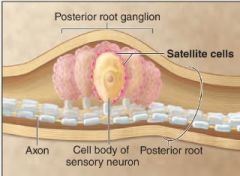
Satellite cells
|
|
|
What is a ganglion?
|
a collection of neuron cell bodies located outside the CNS
|
|
|
_____ physically separate cell bodies in a ganglion from their surrounding interstitial fluid
|

satellite cells
|
|
|
_____ regulate the exchange of nutrients and waste products between neurons and their environment in the PNS
|

satellite cells
|
|
|
What is another name for neurolemmocytes?
|

Schwan cells
|
|
|
What is another name for Schwan cells?
|

neurolemmocytes
|
|
|
These flattened cells ensheathe PNS axons to form a myelin sheath
|
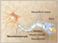
neurolemmocytes
|
|
|
These are large cells with a bulbous body and slender cytoplasmic extensions or processes (CNS)
|

oligodendrocytes
|
|
|
The processes of _____ (CNS) ensheathe portions of axons of many different neurons
|
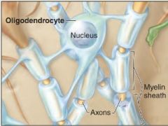
oligodendrocytes
|
|
|
The wrapping around an axon by either an oligodendrocyte or neurolemmocyte
|
myelin sheath
|
|
|
This protective covering around the axon insulates and prevents the passage of ions through the axonal membrane. This allows for faster action potential propagation within the CNS
|
myelin sheath
|
|
|
If a person suffers from meningitis, which type of glial cell usually replicates in response to the infection?
|

Microglia
|
|
|
Which specific type of glial cells ensheaths axons in the PNS?
|

Neurolemmocytes
|
|
|
_____ is the process by which part of an axon is wrapped with myelin.
|
Myelination
|
|
|
_____ is the insulating covering around the axon that consists of repeating concentric layers of plasma membrane of glial cells
|
myelin
|
|
|
Myelination is completed by _____ in the PNS and by _____ in the CNS
|
neurolemmocytes; oligodenrocytes
|
|
|
The high _____ content of the myelin gives an axon a distinct, glossy-white appearance and serves to effectively insulate the axon
|
lipid
|
|
|
The wrapped inner portion of a neurolemmocyte on an axon is called the
|
myelin sheath
|
|
|
The outer-most part of the neurolemmocyte around an axon is made up of _____ and _____ and is called the _____
|
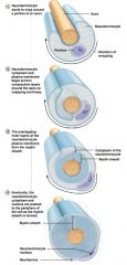
cytoplasm, nucleus; neurilemma
|
|
|
A neurolemmocyte in the PNS can myelinate only a _____ mm portion of an axon
|
1
|
|
|
The gaps between the neurolemmocytes are called _____
|
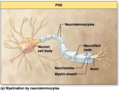
neurofibril nodes
|
|
|
what is another name for a neurofibril node?
|
nodes of Ranvier
|
|
|
What is another name for nodes of Ranvier?
|
neurofibril nodes
|
|
|
What is the difference between what a neurolemmocyte and an oligodendrocite can myelinate?
|
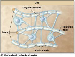
a neurolemmocyte can only myelinate a 1 mm portion of a PNS axon at a time. an oligodendrocyte can myelinate a 1mm portion of multiple axons of the CNS at a time.
|
|
|
Is a neurilemma formed with CNS myelination?
|
No
|
|
|
Is a neurilemma formed with PNS myelination?
|
Yes
|
|
|
Are neurofibril nodes between oligodendrocyte myelin sheaths on an axon?
|

Yes
|
|
|
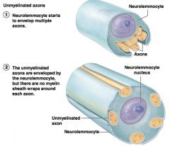
Are all axons myelinated?
|
No
|
|
|
Are PNS unmyelinated axons associated with the neurolemmocyte?
|
Yes, they rest in a depression within the neurolemmocyte, but no sheathe is wrapped around the axon.
|
|
|
Are CNS unmyelinated axons associated with oligodendrocytes?
|
No
|
|
|
What is the function of the myelin sheathe?
|
Insulation
|
|
|
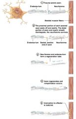
Can a damaged PNS axon regenerate and if so under what conditions?
|
Yes, if the cell body remains intact and a critical amount of neurilemma remains.
|
|
|
The success of PNS axon regeneration depends upon two primary factors:
|
1 - the amount of damage
2 - the distance between the site of the damaged axon and the structure it innervates the possibility of repair decreases with an increase of either of these two factors. |
|
|
______ play an active role in PNS axon regeneration
|

neurolemmocytes
|
|
|
The process of axon regeneration (PNS) follows what stages? (5)
|
|
|
|
What are some of the reasons regeneration of damaged neurons within the CNS is very limited?
|
1 - oligodendrocytes do not release a nerve growth factor and actively inhibit axon growth by producing and secreting several growth-inhibitory molecules. 2 - the large number of axons crowded within the CNS tends to complicate regrowth activities. 3 - both astrocytes and connective tissue coverings may form some scar tissue that obstructs axon regrowth.
|
|
|
Neurons contain _____ proteins for moving substances across the plasma membrane
|
transport
|
|
|
_____ move substances up (against) a concentration gradient, a process that requires energy
|
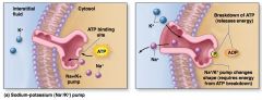
Pumps
|
|
|
Why do pumps require energy?
|

Because they are moving substances against a concentration gradient.
|
|
|
The plasma membrane of neurons contains both ____ pumps and _____ pumps
|
sodium-potassium (Na+/K+)
calcium (Ca2+) |
|
|
_____ provide the means to move a substance down (with) the concentration gradient
|
channels
|
|
|
List the major types of channels (3)
|
Leak (passive) channels
Chemically gated channels Voltage-gated channels |
|
|
What is another name for leak channels?
|

passive channels
|
|
|
What is another name for leak channels?
|
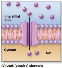
Passive channels
|
|
|
These channels are always open, allowing continuous diffusion of a specific type of ion from a region of high concentration to a region of low concentration.
|

Leak channels
|
|
|
These channels are normally closed and open in response to binding of a neurotransmitter.
|
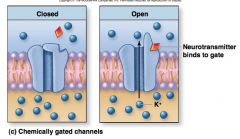
Chemically gated channels
|
|
|
These channels are normally closed and open in response to changes in electrical charge (potential) across the plasma membrane
|

Voltage-gated channels
|
|
|
Most voltage-gated channels have one gate that is in one of two states; open or closed. _____ voltage-gated channels are unique because they have two gates.
|

Na+
|
|
|
What are the two gates of a Na+ voltage-gated channel?
|

activation gate and inactivation gate
|
|
|
What are the activation gate and inactivation gate?
|

The two gates of Na+ voltage-gated channels
|
|
|
What are the three states of voltage-gated Na+ channels?
|

1 - Resting state
2 - Activation state 3 - Inactivation state |
|
|
Describe the resting state of the voltage-gated Na+ channels.
|

Although the inactivation gate is open, the activation gate is closed and entry of Na+ is prevented.
|
|
|
Describe the activation state of the voltage-gated Na+ channels.
|
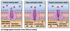
Both the inactivation gate (which remains open) and the activation gate are open (activation gate opens in response to a voltage change); Na+ moves into the cell through the open channel.
|
|
|
Describe the inactivation state of the voltage-gated Na+ channels.
|

Although the activation gate is open, the inactivation gate is TEMPORARILY closed (for several milliseconds) following activation of the Na+ channel - during this time, it cannot be stimulated to reopen, and entry of Na+ is prevented. (The resting state of voltage-gated Na+ channels is reestablished as the inactivation gate opens and the activation closes.)
|
|
|
What pumps and/or channels are located throughout the entire neuron plasma membrane of a neuron?
|

Na+ leak channels, K+ leak channels, and Na+/K+ pumps
|
|
|
These are important in establishing and maintaining the resting membrane potential of neurons.
|
Na+ leak channels, K+ leak channels, and Na+/K+ pumps
|
|
|
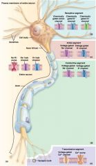
A typical neuron is functionally organized into these four segments:
|

1 - receptive segment
2 - initial segment 3 - conductive segment 4 - transmissive segment |
|
|
T/F Each of the four segments of a typical neuron differs in the primary types of channels and pumps located within its plasma membrane
|

True
|