Reading...
![]()
Play button
![]()
Play button
![]()
Use LEFT and RIGHT arrow keys to navigate between flashcards;
Use UP and DOWN arrow keys to flip the card;
H to show hint;
A reads text to speech;
101 Cards in this Set
- Front
- Back
|
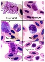
3 types of granulocytes
|
neutrophils, eosinophils, basophils
|
|
|
2 types of agranulocytes
|
monocyte, lymphocyte
|
|
|
characterized by segmented, or lobed nuclei, distinct cytoplasmic granules
|
granulocytic cells
|
|
|
referred to as mononuclear cells, do not have segmented nuclei
|
agranulocytic cells
|
|
|
involved in protecting the body against both infectious disease and foreign invaders
|
immunity
|
|
|
2 types of phagocytic invaders
|
monocytes and neutrophils
|
|
|
3 types of granules that help digest foreign invaders
|
neutrophils, eosinophils, basophils
|
|
|
this type of WBC manufactures proteins and mediators that destroy invaders
|
lymphocytes
|
|
|
what is the most predominant WBC (except in ruminants), mainly phagocytic, involved in inflammation, segmented
|
neutrophil
|
|
|
neutrophils are called what in rabbits, birds, and reptiles
|
heterophils
|
|
|
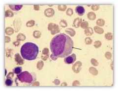
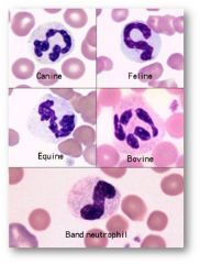
deeply staining, clumped, segmented nuclei with 3-5 lobes. cytoplasm is usually colorless to pale pink in most species or at most are very faint, dusting of tiny granules
|
mature neutrophil morphology
|
|
|
more distinct in nuclear segmentation
|
equine segs
|
|
|
pink to orange cytoplasm
|
bovine seg
|
|
|
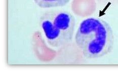
morphology: band shaped nuclei, horseshoe or S shaped. lack segmentation, parallel borders
|
immature neutrophil morphology
|
|
|
may be found normally, in small numbers of domestic animals. indicates release of immature neutrophils, from bone marrow. presence of WBCs in circulation is seen as a result of increased bone marrow activity.
|
band neutrophils
|
|
|
appearance of increased # of band cells and/or more immature forms is referred to as a
|
"left shift"
|
|
|
left shift is generally due to an
|
inflammatory reaction
|
|
|
what are metamyelocytes, myelocytes, promyelocytes, and myeloblasts?
|
stages of maturity for neutrophils
|
|
|
this is often seen with left shift. commonly associated with: inflammation, infection, drug toxicity
|
toxic neutrophilic changes
|
|
|
3 cytoplasmic characteristics of toxic neutrophils
|
Dohle bodies, cytoplasmic vacuolation, toxic granulation
|
|
|
what species show toxic neutrophils during many kinds of illnesses?
|
feline
|
|
|
small, pale bluish-gray irregular inclusions (ribosomal material) in cytoplasm
|
Dohle bodies
|
|
|
Dohle bodies are common in what species? and may be seen with chronic bacterial infection and some viral disease
|
feline
|
|
|
Dohle bodies usually indicate what?
|
mild toxemia
|
|
|
these can be seen in the lymphocyte and neutrophil. associated with septicemia. can also be artifact if sample is held for an extended time in anticoagulant. can range from a few to many. may cause foamy appearance, recorded as moderate to severe
|
cytoplasmic vacuoles
|
|
|
numerous large granules that range in color front dark purple/ red to black. seen in most infectious diseases. may be described by number or by severity. common in horses, rarely seen in other species
|
toxic granulation
|
|
|
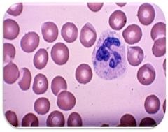
nuclei with five or more lobes. aging change implies a nontoxic environment and prolonged circulation of neutrophils. most frequently seen with steroid use and stress leukograms
|
hypersegmentation
|
|
|
an increase in total number of neutrophils. often seen with inflammatory leukogram
|
neutrophilia
|
|
|
a decrease in circulating neutrophils. may occur as a result of severe inflammation. tissue demand is excessive and exceeds the ability of the bone marrow to supply neutrophils
|
neutropenia
|
|
|
these are necessary for the body to fight serious inflammation or infection
|
neutrophils
|
|
|
functional equivalent of a neutrophil in rabbits, rodents, birds, reptiles, and amphibians. also referred to as pseudoeosinophils. segmented nucleus and variably sized red/brown granules.
|
heterophils
|
|
|
bird granules can be what 2 shapes?
|
oval or needle shaped
|
|
|
much less common than neutrophils (absent or present in very low numbers in normal animals.
|
eosinophils
|
|
|
eosinophil functions to control what 2 things
|
allergic or anaphylactic hypersensitivity
|
|
|
these have a lobulated nucleus and red/orange/pink granules. these contain substances that destroy parasites and others that counteract effects of basophils
|
eosinophils
|
|
|
morphology of eosinophils in cats
|
tiny numerous rod-shaped granules
|
|
|
morphology of eosinophils in dogs/cattle
|
less numerous, round, and vary in size
|
|
|
morphology of eosinophils in horses
|
very large, round and stain bright orange
|
|
|
eosinophils last how many hours in blood stream
|
18-24
|
|
|
this may occur with parasitic disease or allergies
|
eosinophilia
|
|
|
these are involved in allergies and metabolic disorders. relatively rare in blood films. when present, usually occur in association with increased eosinophils
|
basophils
|
|
|
in what species are there few basophilic granules that stain purple to blue-black in basophils
|
canine
|
|
|
in what species are there many round granules that stain light lavenders in basophils
|
felin
|
|
|
in what species are basophils packed with granules and stain dark blue
|
equine/bovine
|
|
|
this is the second highest number of WBC in circulation. most numerous in cattle. grouped into B cells and T cells
|
lymphocytes
|
|
|
B cells in bone marrow are responsible for what
|
humoral immunity. making immunoglobulins
|
|
|
T cells in thymus gland are responsible for what
|
cell-mediated immunity
|
|
|
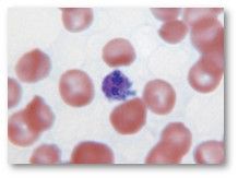
these are small to medium-sized mononuclear cells with a thin rim of light to dark blue cytoplasm and a round, often eccentric nucleus. 7-9um in diameter. cytoplasm may contain azurophilic (blue) granules. components of immune response
|
lymphocytes
|
|
|
cells with dark blue cytoplasm and a darker nucleus. seen in chronic infections. reported as few, moderate, or many
|
reactive lymphocytes
|
|
|
these develop in bone marrow. do not have a function within the bloodstream. circulate briefly before entering the tissue where they become macrophages
|
monocytes
|
|
|
phagocytize large particles and cellular debris that neutrophils cannot handle
|
macrophages
|
|
|
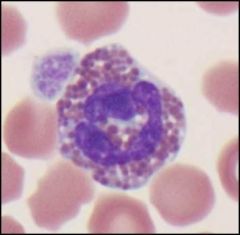
this is a very large WBC with diffuse, less dense nuclear chromatin. nucleus vary in shape. cytoplasm is blue-gray and abundant. nuclear-to-cytoplasm ratio is usually 1:1. vacuoles and/or small fine pink granules may be present
|
monocytes
|
|
|
these may be difficult to differentiate from bands, large lymphocytes or metamyeloctes that are toxic. involved in chronic inflammatory process
|
monocytes
|
|
|
this is caused by increased monocytes. can be seen with viral infections and chronic inflammation disease. (fungal infections or granulomas)
|
monocytosis
|
|
|
largest cell in the bone marrow. unlike other hematopoetic cells, it gets larger as it grows. cytoplasm eventually fragments into multiple irregular pieces that are released to circulation. these fragments are platelets
|
megakaryocyte
|
|
|
how many days does it take to release platelets
|
5 days
|
|
|
these are fragments of megakaryocytic cytoplasm. no nucleus, light granules may be visible. not classified as a cell. round to oval in shape, irregular edges.
|
platelets. aka thrombocytes
|
|
|
what is the normal life span of a platelet
|
10 days
|
|
|
what is the role of a platelet
|
to start clotting process by clumping together to plug damaged vessels
|
|
|
abnormally large platelets can indicate what
|
bone marrow disease or regenerative anemia
|
|
|
blood sample that sits in EDTA for several hours may cause the platelets to do what
|
swell
|
|
|
what are large platelets?
|
young platelets
|
|
|
a decrease in platelet number or function is known as what? it is a condition that affects bone marrow, bacterial and viral infection, immediately following severe hemorrhaging, decreased production of megakaryocytes, platelet destruction, long term antibiotic treatment
|
thrombocytopenia
|
|
|
this is an increase in platelets. response to disease, or following trauma
|
thrombocytosis
|
|
|
this invades platelets causing infectious canine cyclic thrombocytopenia (ICCT). leads to bleeding disorders and fevers in canines; nosebleeds, bleeding under skin. look very similar to platelets
|
anaplasma platys
|
|
|
what vector is anaplasma platys carried by
|
brown tick
|
|
|
what number of platelets seen on a blood smear per 100x field is considered adequate
|
8-10
|
|
|
what are 3 mechanisms of homeostasis
|
vascular response, platelet response, coagulation mechanism
|
|
|
how blood vessels respond to injury
|
vascular response
|
|
|
activation of platelets in clotting
|
platelet response
|
|
|
stimulation of the clotting cascade
|
coagulation mechanism
|
|
|
what is intrinsic
|
intravascular
|
|
|
what is extrinsic
|
tissue system
|
|
|
what 2 organs remove damaged platelets from circulation
|
liver and spleen
|
|
|
7 diseases associated with a secondary bleeding disorder
|
immunologic disease, leukemia, liver disease, splenic disease, uremia, exposure to radiation, DIC (disseminated intravascular coagulation)
|
|
|
this type of test is used to diagnose various bleeding disorders and maintain homeostasis
|
coagulation test
|
|
|
coagulation tests are achieved by 3 interrelationships
|
vascular, platelets, clotting factors
|
|
|
6 types of coagulation tests
|
platelet count; buccal mucosal bleeding time; whole blood clotting time; prothrombin time (PT); partial thromboplastin time (PTT); fibrinogen activity test
|
|
|
normal value for whole blood clotting time for canine? equine?
|
canine: 2-10 min; equine: 4-15 min |
|
|
clotting factors: Factor I: ? Factor II: ? Factor III: ? Factor IV: ? |
I: Fibrinogen; II: Prothrombin; Factor III: Tissue Thromboplastin; Factor IV: Calcium
|
|
|
clotting factors: Factor V: ? Factor VII: ? Factor VIII: ? Factor IX: ? Facotr X: ? |
V: Proaccelerin VII: Proconvertin IX: Christmas factor X: Stuart-Prower factor |
|
|
clotting factors: Factor XI: ? Factor XII: ? Factor XIII: ? |
XI: plasma thromboplastin antecedent XII: Hageman factor XIII: Fibrin-stabilizing factor |
|
|
anaplasma platys
|
|
what species?
|
avian
|
|
|
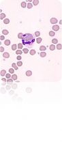
basophils
|
|
|
Dohle bodies
|
|

|
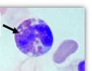
eosinophil
|
|
|
eosinophilic band cells
|
|
|
equine eosinophil
|
|
|
hypersegmented neutrophil
|
|

|
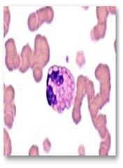
lymphocytes
|
|
|
metamyelocyte
|
|

|
monocyte
|
|
|
myelocyte
|
|
|
neutrophilic band cell
|
|
|
neutrophils
|
|
|
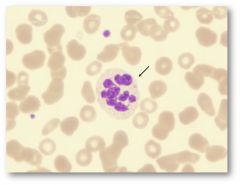
platelets
|
|
|
rabbit heterophil
|
|
|
reactive lymphocytes
|
|
|
toxic granulation
|
|
|
vacuolization
|