Reading...
![]()
Play button
![]()
Play button
![]()
Use LEFT and RIGHT arrow keys to navigate between flashcards;
Use UP and DOWN arrow keys to flip the card;
H to show hint;
A reads text to speech;
63 Cards in this Set
- Front
- Back
|
What is the parietal surface of the spleen in contact with? What ribs of the equine does it course behind?
|
Courses obliquely cranioventrally from the last 3-4 ribs to the ventral third of the 9th-12th intercostal spaces
Parietal surface is in contact with the diaphragm |
|
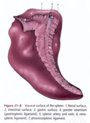
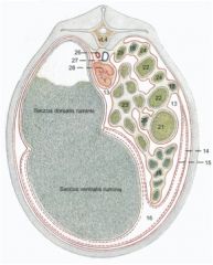
What are 4, 6, and 7 of this equine spleen?
|

Gastrosplenic ligament (4)
Between the hilus of the spleen and greater curvature of the stomach Renosplenic (nephroslplenic) ligament (6) Between the spleen and left kidney Phrenicosplenic ligament (7) Between the spleen and left crus of the diaphragm |
|
|
What is the visceral surface of the ruminant spleen in contact with? What ribs does the dorsal extremity lie under? What are the dorsal and ventral parts attached to? Where is the hilus located?
|
Dorsal extremity lies under the dorsal ends of the last two ribs
Visceral surface is in contact with the left side of the rumen Dorsal part is attached to the left crus of the diaphragm Ventral part is free Hilus is dorsally located on the visceral surface |
|
|
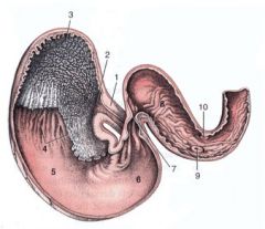
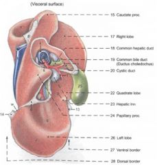
What are the lobes of the equine liver? Where is the gall bladder?
|

Right
Quadrate Left medial Left lateral Caudate lobe with caudate process No gall bladder |
|
|
Where does bile come from in the equine?
|
Bile flows from hepatic ducts to common hepatic duct to major duodenal papilla
|
|
|
Is there a papillary process of the caudate lobe of the equine liver?
|
No
|
|
|
What is embedded in the parietal surface of the equine liver?
|
Caudal vena cava embedded in diaphragmatic surface (another name for parietal surface)
|
|
|
What are the impressions of the visceral surface (5) of the equine liver?
|
Hilus/porta – portal v., hepatic a., hepatic nn., common hepatic duct, lymphatic vessels
Gastric impression Duodenal impression Colic impression -In contact with the diaphragmatic flexure and right dorsal colon Cecal impression -In contact with the cranial part of the base of the cecum |
|
|
What are the impressions of the dorsal border (2) of the equine liver?
|
Renal impression
-In contact with the right kidney Esophageal impression |
|
|
Where are the triangular ligaments of the equine liver found?
|

Right – between right liver lobe and costal part of the diaphragm
Left – between left liver lobe and central tendon of diaphragm |
|
|
What does the coronary ligament connect? Where does it course between?
|
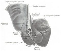
Connects liver to diaphragm
Courses between triangular ligaments |
|
|
Where does the falciform ligament of the equine liver attach? What does it contain? Where does it extend to?
|
Attaches the quadrate and left
medial liver lobes to the sternal part of the diaphragm and floor of abdomen Contains the round ligament of the liver extends to the umbilicus |
|
|
Where does the hepatorenal ligament of the equine liver attach?
|
Attaches caudate process to
right kidney and base of cecum |
|
|
What are the boundaries for liver biopsy in the horse?
|
Right side
Line between tuber coxae and olecranon Line between tuber coxae and point of shoulder Between ribs 11 and 14 |
|
|
To which side is the bovine liver displaced?
Where are its lobes located? |
Liver displaced to the right of the median plane - the whole liver has just been shifted to the right.
Right lobe is caudodorsal Left lobe is cranioventral |
|
|
What are the lobes of the bovine liver?
|
Right
Quadrate Left Caudate with caudate and papillary processes |
|
|
What lobes of the liver does the bovine gall bladder course between?
|
Gall bladder is located between the right and quadrate lobes
|
|
|
What is the ligament of the diaphragmatic surface of the bovine liver?
|

Falciform ligament (13) is attached to the diaphragmatic surface from the esophageal impression to the notch for the round ligament of the liver
|
|
|
What is the area attached to the diaphragm devoid of serous covering of the bovine liver?
|

(16)
The area nuda |
|
|
What are the ligaments of the ruminant liver?
|
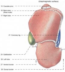
Triangular ligaments
Right (7) – between right liver lobe and dorsal abdominal wall Left (14) – between esophageal impression and diaphragm ventral to the esophageal hiatus Coronary ligament (21) Connects liver to diaphragm Courses between triangular ligaments Falciform ligament (13) Attached to the diaphragmatic surface of liver from esophageal impression to the notch for the round ligament of the liver – courses to the diaphragm Contains the round ligament of the liver Hepatorenal ligament Attaches caudate process to the ventral surface of the right kidney |
|
|
What makes up the bile system of the bovine liver?
|
Gall bladder
Hepatic ducts Cystic duct Bile duct |
|
|
What are the Boundaries for liver biopsy in the ox?
|
Right side
Intersection of the 11th intercostal space and a line drawn from the olecranon to the tuber coxae |
|

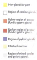
What are the glandular distributions of the bovine stomach?
|
Rumen, reticulum and omasum are nonglandular
|
|
What are the glandular distributions of the equine stomach?
|
see
|
|
|
What is the capacity of the equine stomach?
|
5-15 L
|
|
|
What is the parietal surface of the equine stomach in contact with?
What about the visceral surface? |
Parietal surface
In contact with the diaphragm and liver Visceral surface In contact with the terminal part of the ascending colon, pancreas, descending colon, small intestine and greater omentum |
|
|
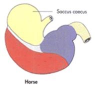
What is the area dorsal to the cardia of the equine stomach called?
|
fundus or saccus cecus.
|
|

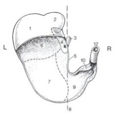
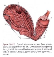
Name these equine stomach areas.
|
1. Esophagus
2. Cardia 3. Fundus (saccus cecus, blind sac) 4. Margo plicatus (marginal fold) 5. Body 6. Pyloric region 7. Pylorus |
|
|
What are the gastric ligaments in the horse?
|
Gastrophrenic ligament
Connects greater curvature of the stomach from the cardia to the left side with the crura of the diaphragm. Gastrosplenic ligament Courses from the left region of the greater curvature of the stomach to the hilus of the spleen Is continuous ventrally with the greater omentum |
|
|
What are the anatomical reasons why it is difficult for the horse to vomit? (5)
|
Tunica muscularis in the distal 1/5 of the esophagus is twice as thick as in other regions of the esophagus forming a strong lower esophageal sphincter.
Tunica muscularis in distal 1/5 of esophagus is entirely smooth muscle and is, therefore, not under voluntary control. Esophagus enters stomach at an oblique angle. When pressure within the stomach increases, the cardia closes tighter. Stomach does not touch the ventral abdominal wall. It is situated dorsal to the ventral and dorsal portions of the ascending colon. Abdominal muscular contractions associated with vomiting will first be absorbed by the ascending colon. Saccus cecus and pyloric antrum are positioned cranial to the cardia. Gastric contractions are more likely to force contents into these two regions rather than through the cardia. |
|
|
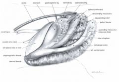
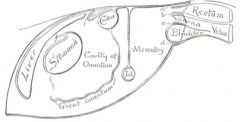
What does the greater omentum of the horse connect?
What is the omental bursa? Where are the boundaries of the epiploic foramen? What's the clinical signifigance of this foramen? |

Greater omentum
-Connects the greater curvature of the stomach and the initial part of the duodenum with the terminal part of the large colon and initial part of the small colon (which connect to the dorsal body wall) Omental bursa -Located between the superficial and deep layers of the greater omentum Epiploic foramen -Dorsal boundary – caudate process of liver and caudal vena cava -Ventral boundary – pancreas, hepatoduodenal ligament and hepatic portal vein -Small Intestine could go through this foramen into the bursa |
|
|
What does the lesser omentum in the horse connect? What are its ligaments?
|
Connects the lesser curvature of the stomach and the first part of the duodenum with the liver
Hepatogastric ligament and hepatoduodenal ligament |
|

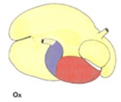
Name A, B, C, D
|
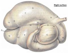
A. Rumen
B. Reticulum C. Omasum D. Abomasum |
|
|
What are the capacities of the ox and small ruminant stomachs?
|
Ox - 60L or more
Small ruminant - 15-18 L |
|
|
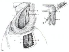
What are the three parts of the gastric groove?
|
The reticular groove (25) is located between the cardia (24) and the reticulo-omasal orifice (28)
The omasal groove is located between the reticulo-omasal and omasoabomasal orifices Abomasal groove |
|

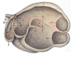
What are 1-6?
|
1. Left lip of the reticular groove
2. Right lip of the reticular groove 3. Cardia 4. Reticulo-omasal orifice 5. Wall of reticulum 6. Ruminoreticular fold |
|
|
What are the intraruminal and ruminoreticular orifices of the bovine rumen?
|
Intraruminal orifice – opening between dorsal and ventral sacs of the rumen
Ruminoreticular orifice – opening between rumen and reticulum |
|
|
What is the area of the rumen that the reticulum opens into? How is it different from the rumenoreticular orifice?
|
atriumventriculi - which is the general dorsal region between the rumen and reticulum
The rumenoreticular orifice is the entire opening. |
|
|
Where is the insula of the bovine rumen?
|
Between right accessory pillar and right longitudinal pillar
|
|
|
What are the four ways in which the small ruminant rumen differ from the large animal rumen?
|
Sheep and goat – ventral sac is relatively larger and extends more to the right of the median plane than in the ox
Caudoventral blind sac more extensive than caudodorsal blind sac Left longitudinal groove does not connect with caudal groove The dorsal coronary grooves are very short or absent |
|
|
What is the level to find the reticulum? To which side of midline does it lie?
What do the parietal and visceral surfaces of the reticulum contact? |
Located between 6th and 7th or 8th ribs (just caudal to the diaphram at the 6th rib)
Lies mostly to the left of midline Parietal surface is in contact with the diaphragm and liver Visceral surface is in contact with the rumen |
|
|
Is the reticulum relatively larger in the goat and sheep or ox?
|
The reticulum in the sheep and goat is relatively larger than in the ox
|
|
|
Why is the reticulum honeycombed?
|
Traps particles that are too small to be regurgitated and need to just go on to the omasum.
|
|
|
How many ruminal and reticular contractions per minute?
What are fewer/more per minute called? |
1-3
Less – ruminal atony More – hypermotility |
|
|
What stimulates the growth of ruminal papillae?
|
VFAs
|
|
|
Which flank can ruminal contractions be seen in?
|
Left
|
|
|
At which rib space are reticular contractions?
How many contractions should you feel per minute at the paralumbar fossa? |
Stick hand in paralumbar fossa, should feel 2 contractions per minute
Reticular contractions – left 6th-7th IC space |
|
|
At what level is the omasum located in the ox? What are the parietal and visceral surfaces in contact with?
|
Located mainly to the right of midline, opposite the 7th-11th ribs
Parietal surface is in contact with the diaphragm, liver and body wall (7th-9th intercostal spaces) Visceral surface is in contact with the rumen, reticulum and abomasum |
|
|
Is the omasum smaller or larger in the sheep and goat compared to the ox? What level is it located at?
|
Omasum in the sheep and goat is much smaller than the ox; located at 9th-10th ribs and has no contact with the body wall
|
|
|
What is the function of the omasum?
|
The omasum is thought to act as a pump transferring material from the reticulum to the abomasum as well as aiding in absorption
|
|
|
What do the visceral and parietal walls of the abomasum contact?
What region is the xiphoid region in? What does the body extend between? |
Parietal surface is in contact with the abdominal floor
Visceral surface is in contact with the rumen and omasum Fundus is in the xiphoid region Body extends caudally between ventral sac of the rumen and the omasum lying toward the left of midline |
|
|
At what level is the pylorus of the abomasum located?
What is attached to the lesser curvature of the abomasum? Is the abomasum relatively larger or smaller than that of the ox? |
Pylorus is at the ventral end of 9th-10th intercostal space
Greater curvature is attached to the superficial layer of the greater omentum Lesser curvature is attached to the lesser omentum In the sheep and goat, the abomasum is relatively larger and longer than in the ox |
|
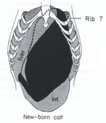
How does the bovine abomasum change by age?
What is a common abomasomal ailment? |
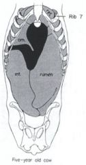
Newborn calf abomasum are huge, soon, it shrinks down. Here we see a bovine at 5 with the abomasum in a normal position with the base pushed to the left by the omasum.
The abomasum can get displaced to the left (more common) or right. |
|

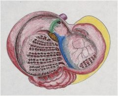
What abnormality is this?
|
left displacement of the abomasum
|
|
|
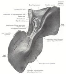
What is the epiploic foramen of the ruminant? What are its boundaries?
|
Entrance into omental bursa (16) which is located between superficial (14) and deep (15) layers of the greater omentum
Lateral boundary – caudate process of the liver Medial boundary – mesoduodenum and pancreas Dorsal boundary – caudal vena cava Ventral boundary – hepatoduodenal ligament |
|
|
Where does the lesser omentum of the ruminant course between?
|
Courses between the lesser curvature of the abomasum and liver
|
|
|
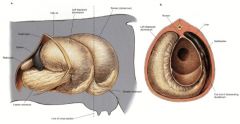
What are the compartments of the camelid stomach? What percentages of the stomach contents do they hold?
|
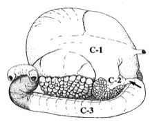
(right lateral view shown)
C 1 (cranial sac and caudal sac) Largest compartment, 83% of contents, fermentation vat, saccules. C 2 Holds 6% of contents Mixing and fermentation, saccules C 3 Holds 11% of contents Initial 4/5 – similar to the saccules of C1 and C2 Distal 1/5 – acid secretion |
|
|
What are a special feature of the ventral portions of C1 and C2 of the camelid stomach? What is their function?
|
Saccules in the ventral portions of C1 and C2
Contain glands that secrete bicarbonate to buffer contents of C1 and C2 and maintain bacteria and protozoa |
|
|
Is the camelid a ruminant?
|
A pseudo-ruminant
|
|
|
What is 10 here of the porcine stomach? It's a unique feature of the porcine stomach.
|
Torus pyloricus
It's a unique feature of the porcine stomach. |
|
|
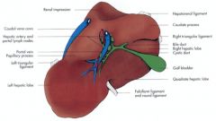
What are the impressions of the visceral surface of the bovine liver?
|
Visceral surface
Hilus/porta – portal v., hepatic a., hepatic nn., hepatic duct, lymphatic vessels Omasal impression (q) Reticular impression (r) Abomasal impression |
|
|
What are the impressions of the right, ventral and dorsal borders of the bovine liver?
|

Right border
Impression for right kidney (v) and adrenal gland Ventral border Gall bladder fossa (more distinct in sheep and goat) Notch for round ligament (p) Dorsal border Groove for caudal vena cava (h) Esophageal impression (w) |
|

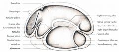
What are 1, 3, 4, 5, 6, 7?
|
Omasoabomasal opening (1)
Fundus (3) Body (4) Pyloric region (5) Torus pyloricus (6) Pylorus (7) |