Reading...
![]()
Play button
![]()
Play button
![]()
Use LEFT and RIGHT arrow keys to navigate between flashcards;
Use UP and DOWN arrow keys to flip the card;
H to show hint;
A reads text to speech;
38 Cards in this Set
- Front
- Back
|
Neurofibroma
You would need to resect that part of the nerve which would probably cause some deficits |
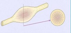
What type of tumor does this exemplify? What kind of treatment would be needed?
|
|
|
Schwannoma
Tumor could probably be teased off from the nerve, leaving it intact |
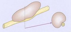
What type of tumor does this exemplify? What type of treatment would this need?
|
|
|
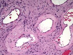
Unencapsulated, but well-circumscribed masses of spindle cells, which occur in the dermis (cutaneous), in the peripheral nerve (solitary), or in a large nerve trunk (plexiform)
|
Neurofibromas
|
|
|
What type of Neurofibroma is associated with NF-1? What may this transform into?
|
Plexiform
Malignant Peripheral Nerve Sheath Tumor (MPNST) |
|
|
Neurofibromas
|
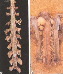
What are seen in these pictures?
|
|
|
A moderately firm, usually benign, unencapsulated, slow-growing heterogeneous tumors of the nervous system arising from the supporting cells (Schwann cells) of peripheral nerves
|
Neurofibroma
|
|
|
How are Schwannoma's & Neurofibromas different?
|
In contrast to Schwannomas - another type of tumor arising from Schwan cells - Neurofibromas incorporate all sorts of cells and structural elements in addition to the Schwann cells
|
|
|
What syndrome are Schwannomas associated with?
|
Neurofibromatosis-2
|
|
|
Where do Schwannomas typically occur?
|
1. CN VIII = Vestibular branch = Acoustic neuroma
2. CN V (Trigeminal) 3. Spinal nerve roots 4. Peripheral nerves |
|
|
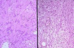
What is the pathology seen in Schwannomas?
|
Encapsulated
Micro: -Antoni A = interlacing bundles of elongated cells with palisading nuclei -Antoni B = looser, less cellular pattern |
|
|
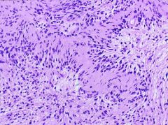
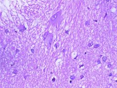
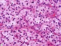
Schwannoma
|
What is this seen in?
|
|
|
These are the classic microscopic appearances of a schwannoma, which is benign. Note the more cellular "Antoni A" pattern on the left with palisading nuclei surrounding pink areas (Verocay bodies). On the right is the "Antoni B" pattern with a looser stroma, fewer cells, and myxoid change
|
What is this showing?
|
|
|
Autosomal dominant disorder that is caused by a mutation of a tumor suppressor gene located on chr. 17
|
Neurofibromatosis-1
|
|
|
What are the clinical features of Neurofibromatosis-1?
|
1. Neurofibromas
2. Gliomas of the Optic nerve 3. Lisch nodules = pigmented nodules of the iris 4. Cafe-au-lait spots |
|
|
A 14-year-old girl presents to your clinic complaining of multiple nodules on her skin. She tells you that her mother suffers from a similar condition. Upon examination, you find multiple coffee-colored macules on her torso & limbs & pigmented nodules on her irises. You suspect her condition is due to an Autosomal Dominant genetic disorder. Dx?
|
Neurofibromatosis-1
|
|
|
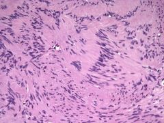
Neurofibroma in a NF-1 patient
|
What is this showing?
|
|
|
Malignant peripheral nerve sheath tumors (MPNST) that arose from from a Neurofibroma
|

What is this showing?
|
|
|
Syndrome in which the normal product is Neurofibromin, a tumor suppressor gene....but is mutated
|
NF-1
|
|
|
What are the clinical manifestations of NF-2?
|
1. Bilateral Acoustic Schwannomas
2. Multiple Meningiomas 3. Gliomas 4. Hamartomas |
|
|
Rare (1/40,000) Autosomal Dominant disorder that is due to a mutation on chr. 22 in which a tumor suppressor gene, Merlin, is mutated
|
NF-2
|
|
|
Schwannoma in NF-2
|
What is seen here?
|
|
|
Autosomal dominant disorder that is due to either a mutation of the Hamartin gene on chr. 9 or the Tuberin gene on chr. 16
|
Tuberous Sclerosis
|
|
|
Disorders that has Hamartomas in the following areas:
-Cysts = liver, kidneys, pancreas -Myomas = heart, lungs -Cutaneous = angiofibromas, shagreen patches, ash-leaf spots, subungal fibromas |
Tuberous Sclerosis
|
|
|
Disorder that has Hamartomas in the CNS that include Cortical Tubers (neurons & cells of intermediate phenotype) & Subependymal Nodules (candle guttering)
|
Tuberous Sclerosis
|
|
|
Tuberous Sclerosis
|
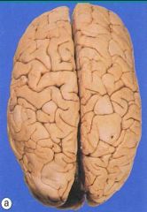
What disorder is this?
|
|
|
Tuberous Sclerosis
-subependymal nodules = hamartomas |
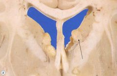
What disorder is this?
|
|
|
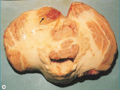
Disorder that has a neoplasm called a Subependymal Giant Cell Astrocytoma
|
Tuberous Sclerosis
|
|
|
Tuberous Sclerosis
|
What disorder is this?
|
|
|
Mental retardation + Hamartomas in the brain & kidneys
|
Tuberous Sclerosis
|
|
|
What symptoms does Tuberous Sclerosis cause?
|
Mental Retardation & Seizures
Hypopigmented skin lesions = "ash leaf" lesions Angiolipomas in the kidneys |
|
|
Autosomal dominant syndrome that has a mutation of a tumor suppressor gene on chr. 3
|
von Hippel-Lindau
|
|
|
Renal cell CA + Cavernous Hemangiomas + Adenomas = ?
|
von Hippel-Lindau
|
|
|
Where do Hemangioblastomas occur in von Hippel-Lindau syndrome?
|
1. Cerebellum
2. Retina 3. Brain stem 4. Spinal Cord |
|
|
Hemangioblastoma of the Cerebellum with Retinal Angiomas = ?
|
von Hippel-Lindau
|
|
|
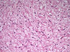
What can Hemangioblastoma's in von Hippel-Lindau syndrome produce?
|
EPO -> polycythemia
|
|
|
Hemangioblastoma in von Hippel-Lindau syndrome
|
What is seen here?
|
|
|
Hemangioblastoma in von Hippel-Lindau
-foamy cells & high vascularity |
What syndrome is this seen in?
|
|
|
Neurofibromatosis-1
-Lisch nodules Neurofibromas Gliomas of the Optic Nerve Cafe au lait spots |
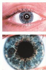
What syndrome is this seen in?
What other clinical manifestations does this syndrome have? |