Reading...
![]()
Play button
![]()
Play button
![]()
Use LEFT and RIGHT arrow keys to navigate between flashcards;
Use UP and DOWN arrow keys to flip the card;
H to show hint;
A reads text to speech;
27 Cards in this Set
- Front
- Back
|
hilium
|
location on kidney where ureters, blood vessels, nerves and lymphatics pass
|
|
|
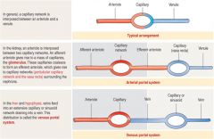
ARTERIAL PORTAL SYSTEM - KIDNEY
VENOUS PORTAL SYSTEM – LIVER & PITUITARY |
|
|
A coronal view (left) shows the major blood vessels of the kidney. The microvascular components extending into the cortex and medulla from the interlobular vessels are shown on the right. Pink boxes indicate vessels with arterial blood and blue indicate the venous return. The intervening lavender boxes and vessels are intermediate sites where most reabsorbed material re-enters the blood.
|
|

efferent arterioles of cortical nephrons
|

give rise to plexus of peritubular capillaries
|
|
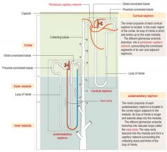
efferent arterioles of juxtamedullary nephrons give rise to
|

vasa rectae spuria projecting radially
toward hilum. |
|
|
afferent vs efferent tubular size
|
efferent arteriole is smaller than afferent; creates pressure to drive filtration process; regulated by arteriole smooth muscle cells
|
|

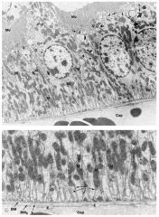
function of endothelial fenestrations
|
permeable to H20, Na, urea, glucose and small proteins (<60 kD); endothelial cells negatiely charged (heparan sulfate -> slows passage of anionic proteins)
|
|
|
mutation in nephrin gene
|
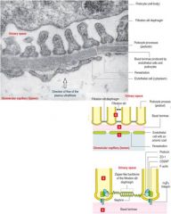
Podocyte pedicels sit on GBM and form filtration slits. Filtration slit
diaphragm is composed of nephrin. Mutation in nephrin gene is cause of congenital nephrotic syndrome (massive proteinuria). |
|
|
mesangial cell
|
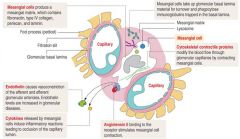
provide phagocytosis, structural support, and secret IL-1 and PDGF in response to glomerular injury
|
|
|
Goodpasture syndrome
|
autoimmune disorder
progressive glomerulonephritis and pulmonary hemorrhage due to anti-collagen a3(IV) antibodies binding to and disrupting glomerular and alveolar basal laminae. GBM is produced by both endothelial cells and podocytes: Type IV collagen, laminin, fibronectrin an dproteoglycans (enrinched in GAGS via heparan sulfate) |
|
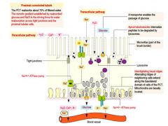
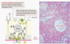
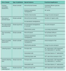
Proximal convoluted tubule
|
receives ultrafilate from urinary space of Bowmans capsule
is the initial and MAJOR site of absorption note brush border |
|
|
loop of henle
|
|
|
distal convoluated tubule
|
|
|
macula densa cells
|
chemoreceptors
feedback system low Na+ => ATP, ACe in endothelial cells system bp = renin - angiotensin - aldosterone |
|
|
ADH
|
regulates water permeability in collecting ducts
CDs also secrete H+ and bicarb -> important for blood pH balance |
|
|
principal cells in collecting tubules
|
reabsorb Na and H20
|
|
|
intercalated cells
|
secrete H+/HC03
|
|
|
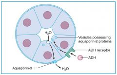
Diagram of the induction of permeability to water in a collecting tubule. When
antidiuretic hormone (ADH) binds to basal receptors of collecting tubule cells, it triggers the movement of cytoplasmic vesicles to the cell surface. These vesicles, containing aquaporin-2 water transporters, allow the entry of water, which then exits the cell at its basolateral surface via aquaporin-3 and -4 channels. |
|
|
EPO
|
EPO is produced mainly by peritubular fibroblasts in the cortex)
|
|
|
Renin
|
(Renin is produced by juxtaglomerular cells in afferent arteriole)
|
|
|
atrial natriuretic peptide
|
intercalated cells of collecting ducts;
production much lower than in atrial cardiomyocytes) |
|
|
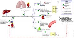
renin angiotensin system
|
|
|
renal papilla
|
|
|
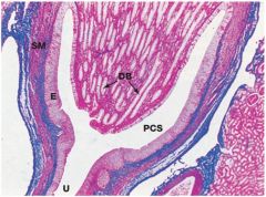
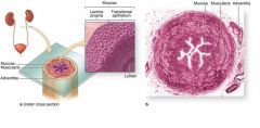
Each ureter carries urine from the renal pelvis of the kidney to the urinary bladder for storage prior to voiding via the urethra.
(a): Diagram of a ureter in cross section shows a characteristic pattern of longitudinally folded mucosa, surrounded by a thick muscularis that moves urine by regular waves of peristalsis. The lamina propria is lined by a unique stratified epithelium called transitional epithelium or urothelium resistant to the potentially deleterious effects of contact with hypertonic urine. (b:) Histologically the muscularis is much thicker than the mucosa and an adventitia is also present. [H&E]. |
|
|
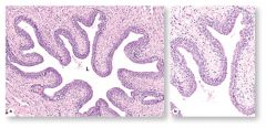
The urethra is a fibromuscular tube that carries urine from the bladder to the exterior of the body.
(a): A transverse section shows that the mucosa has large longitudinal folds around the lumen (L) [H&E]. (b): A higher magnification of the urethral epithelium is shown in this micrograph. The thick epithelial lining is stratified columnar in some areas and pseudostratified columnar elsewhere, but becomes stratified squamous at the distal end of the urethra [H&E]. |
|
|
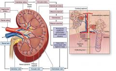
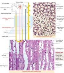
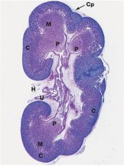
general anatomic and histologic organization of the kidney
|
|
|

compound tubular gland covered with thin capsule of dense connective tissue & fat
parenchyma: outer dark red cortex, striated medulla, funnel-shaped pelvis medulla composed of 12 -15 renal pyramids with apex of papilla extending into calyx hilum on medial surface: ureters, blood vessels, nerves, lymphatics pass |