Reading...
![]()
Play button
![]()
Play button
![]()
Use LEFT and RIGHT arrow keys to navigate between flashcards;
Use UP and DOWN arrow keys to flip the card;
H to show hint;
A reads text to speech;
17 Cards in this Set
- Front
- Back
|
Bones of the shoulder girdle - function
|
Clavicle and scapula - provide a mobile base to attach the limbs to the trunk
|
|
|
Functions of the clavicle (3)
|
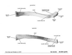
- Strut to distance the scapula and upper limb from the chest wall
- Articulates with the sternum and the acromion process of the scapula - Transmits forces to the axial skeleton (fracture - fall on an outstretched arm) |
|
|
Scapula - articulation and function
|
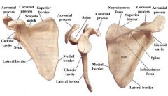
- Articulates with the humerus at the glenoid fossa
- Provides large, flat surfaces and rough processes for muscle attachment |
|
|
Sternoclavicular joint features
|
Between - medial end of clavicle and upper lateral edge of manubrium
Atypical synovial joint - surfaces covered by fibrocartilage Enclosed by synovial capsule, reinforced by anterior and posterior sternoclavicular and costoclavicular ligaments Joint divided into two cavities by a fibrocartilaginous disc |
|
|
Acromioclavicular joint features
|
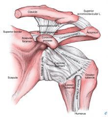
Between lateral end of clavicle and medial edge of acromion process
Atypical synovial joint Weakly encapsulated - stabilised by coracoclavicular and acromioclavicular ligaments |
|
|
Glenohumeral joint features
|
Between glenoid fossa and humeral head
Most mobile joint in body - ball-and-socket synovial joint Shallow joint - low surface area of articulation (unstable - prone to dislocation) Thin synovial capsule - loose inferiorly to permit wide range of movement Capsule strengthened by rotator cuff tendons Glenoid labrum (fibrocartilaginous rim at fossa margin) deepens the joint and helps to stabilise |
|
|
Bones of the upper arm
|
- Humerus
- Radius - Ulna |
|
|
Humerus - features and functions
|
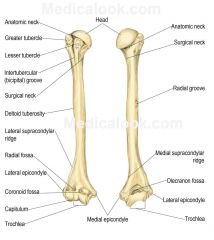
Mobile lever - directs forearm movement in any direction
Articulates the scapula at the head, the radius at the capitulum and the ulna at the trochlea. Forearm angled laterally with respect to the humerus (carrying angle) by the trochlea (projects further at its medial that its lateral border) Often fractures at the 'surgical neck' |
|
|
Radius - features and functions
|

Thick, disc-like head articulates with humerus
Distal end articulates with scaphoid and lunate carpal bones Colles fracture - fractured radius due to fall on an outstretched hand |
|
|
Ulna - features and functions
|
Articulates with humerus at the trochlea notch via the olecranon which prevents overextension of (locks) the elbow joint
Articulates with the radial head at the radial notch |
|
|
Elbow joint features
|
Synovial hinge joint
Articulations - trochlea of humerus and trochlea notch of ulna, humerus capitulum and radial head, radial head and radial notch (proximal radio-ulna joint) Capsule covers all 3 articulations - reinforced by radial and ulna collateral ligaments (lateral and medial ligaments) Proximal radio-ulna joint held together by the annular ligament - permits rotation of the radial head on ulna CLINICAL: fall on an outstretched hand = posterior dislocation |
|
|
Interosseous membrane
|
Joins shafts of radius and ulna
Fibrous sheet - syndesmosis |
|
|
Distal radio-ulna joint
|
Between ulnar notch of radius and the ulnar head
Radius and ulna united by intra-articular disc - a triangle of fibrocartilage |
|
|
Carpal bones - features and functions
|

Transversely arched - creates a hollow
Provides flexible yet firm basis on which muscles can exert their action Scaphoid prone to fracture through fall on outstretched hand |
|
|
Radio-carpal joint
|
Synovial ellipsoid joint between distal end of radius + intra-articular disc and the proximal row of carpals
Strengthened by radial and ulnar collateral and dorsal and palmar ligaments Flexor retinaculum (transverse carpal ligament) - runs from (medially) pisiform and hook of hamate to (laterally) scaphoid and trapezium Forms the carpal tunnel - the flexor tendons of the digits and the median nerve run through this Merges with palmar aponeurosis (protective, tendon-like sheet) originating from the tendon of palmaris longus Extensor tendons run under the thinner, extensor retinaculum - runs across the back of the wrist from the pisiform and hook of hamate to the radius forming channels by spanning grooves on the dorsal aspect of the distal radius. |
|
|
Metacarpals - features and functions
|

1st is support for the base of the thumb
Remaining 4 form the framework of the palm Relatively immobile (only the thumb moves freely) |
|
|
Phalanges - features and functions
|
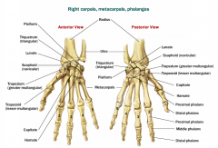
Bones of the fingers (3) and thumb (2)
Function as a unit rather than individual bones |