Reading...
![]()
Play button
![]()
Play button
![]()
Use LEFT and RIGHT arrow keys to navigate between flashcards;
Use UP and DOWN arrow keys to flip the card;
H to show hint;
A reads text to speech;
45 Cards in this Set
- Front
- Back
|
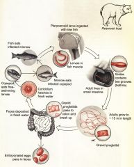
Life cycle of Diphyllobothrium latum
|
|
|

how long can this grow?
|
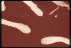
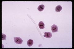
Gravid proglottids, Diphyllobothrium latum
grows up to 10 meters long with 3-4,000 proglottids |
|
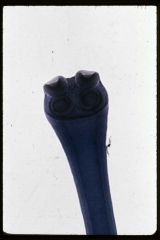
identify the scolex, what characteristic does it have?
|
Scolices of Diphyllobothrium latum
the scolex is spatulate with groves called bothridia |
|
|
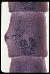
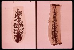
Gravid proglottid of Diphyllobothrium latum
|
|
|
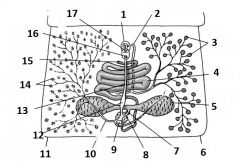
Gravid proglottid of Diphyllobothrium latum
1. Cirrus pouch 2. vas deferens 3. testes 4. uterus 5. ovary 6. Oocapt 7. Oviduct 8. Mehlis' gland 9. Vitelline duct 10. Seminal receptacle 11. vagina 12. vitelline follicles 13. uterine pore 14. vaginal pore 15. male genital pore |
|

|
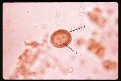
Eggs of Diphyllobothrium latum
1. Abopercular thickening of egg shell |
|

|
Egg of Diphyllobothrium latum
|
|
|
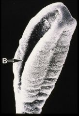
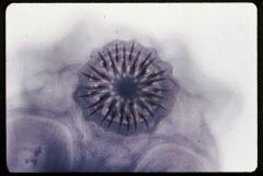
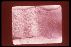
SEM of Diphyllobothrium latum scolex shoowing botridium
|
|
|
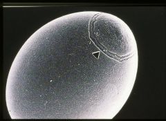
SEM of Diphyllobothrium latum egg showing edges of operculum
|
|
|
Taenia pisiformis life cycle
|
|
|
|
Scolex of Taenia pisiformis
|
|
|
Mature proglottid of Taenia pisiformis
|
|
|
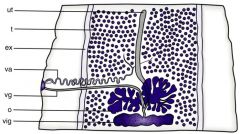
Mature proglottid of Taenia pisiformis anatomy
ex: excretory canal o: ovary t: testis ut: uterus va: vas deferens vg: vagina vig: vitelline gland |
|
|
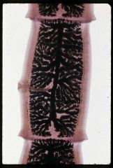
Gravid proglottid of Taenia pisiformis
|
|
|
Egg of taeniids. Species can’t be identified beyond family (Taeniidae) on basis of egg morphology
1. gelatinous granular layer 2. striated 3. inner 4. embryophores 5. capsule |
|
|
Typical taeniid egg in stool sample
1. gelatinous layer 2. embryophore 3. oncosphere, hexacanth |
|
|
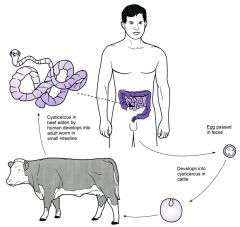
Taenia saginata, life cycle
|
|
|
|
Strobila of Taenia saginata
|
|
|
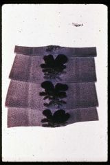
Gravid proglottids, Taenia saginata in stool sample
1. lateral genital pores |
|

|
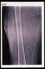
Taenia saginata strobila, only “part” of tapeworm recovered after vermifuge
|
|
|
Scolex of Taenia saginata
|
|
|
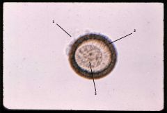
Taenia Eggs
1. embryophore 2. oncosphere with hooks |
|
|
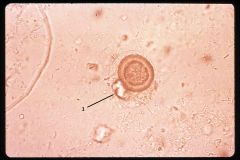
Taenia sp. egg in fecal sample
1. gelatinous matrix |
|
|
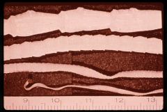
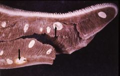
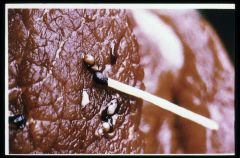
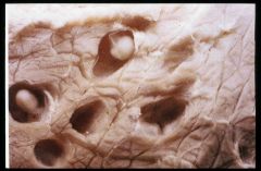
Cysticerci (arrows) of Taenia saginata in cow (10 years old) tongue
|
|
|
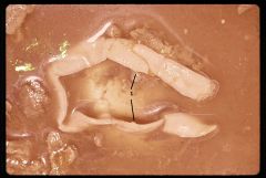
Cysticercus of Taenia saginata in a beef cut
|
|
|
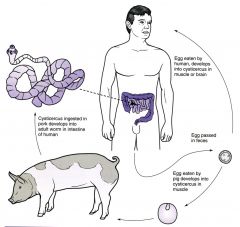
Taenia solium life cycle
|
|
|
|
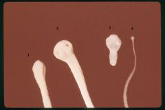
Tapeworm scolices, comparison view
1. Diphyllobothrium latum 2. Taenia saginata 3. Taenia solium 4. Hymenolepis nana |
|
|
Scolex of Taenia solium, “Pork Tapeworm”
|
|
|
Comparison of Taenia gravid proglottids from humans
|
|

|
Cysticercus of Taenia solium
1. scolex |
|
|
Cysticerci, Taenia solium, in pig muscle
|
|

|
Cysticercosis, subcutaneous
|
|
|
Calcified cysticerci
|
|
|
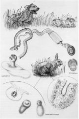
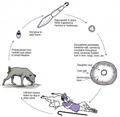
Pastoral life cycle of Echinococcus granulosus
|
|
|
|
Echinococcus granulosus in dog small intestine
|
|

|
Echinococcus granulosus, adult tapeworm
|
|
|
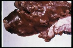
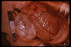
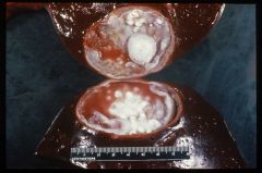
Multiple hydatid cysts in sheep liver
|
|
|
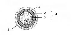
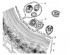
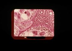
Morphology of the hydatid cyst, Echinococcus granulosus
1. Brood capsule 2. protoscolices 3. germinal layer 4. laminated cuticular layer 5. outer fibrous layer |
|

|
Morphology of the hydatid cyst, Echinococcus granulosus
1. Germinal layer 2. brood capsule 3. protoscolex arrow: extend of the laminated layer |
|
|
Morphology of the hydatid cyst, Echinococcus granulosus
1. fibrous layer limits |
|
|
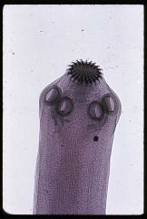
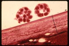
Protoscolices of Echinococcus granulosus
|
|

|
Hydatid cyst in human lung
|
|
|
Hydatid in pre-teen immigrant in NYC
|
|
|
Hydatid in human liver detected during post-mortem
|
|
|
Echinococcus multilocularis, alveolar cyst
|