![]()
![]()
![]()
Use LEFT and RIGHT arrow keys to navigate between flashcards;
Use UP and DOWN arrow keys to flip the card;
H to show hint;
A reads text to speech;
187 Cards in this Set
- Front
- Back
- 3rd side (hint)
|
central nervous system |
the complex of nerve tissues that controls the activities of the body; consists of the brain and spinal cord |
|
|
|
neural tube |
the embryonic structure that becomes the brain and spinal cord |
|
|
|
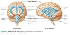
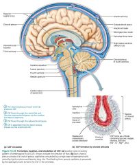
ventricles |
fluid-filled spaces within the brain; lined by ependymal cells |

|
|
|
1. lateral ventricles (x2) 2. third ventricle 3. fourth ventricle |
3 sets of brain ventricles |

|
|
|
lateral ventricles |
large C-shaped brain ventricles; chambers that reflect the pattern of cerebral growth (like a ram's horns) |

|
|
|
septum pellucidum |
thin median membrane separating the right and left lateral ventricles |

|
|
|
interventricular foramen |
channel connecting the lateral ventricles to the third ventricle |

|
|
|
third ventricle |
narrow brain ventricle in the diencephalon |

|
|
|
cerebral aqueduct |
canal-like duct connecting the third and fourth ventricles, runs through the midbrain |
|
|
|
fourth ventricle |
large brain ventricle dorsal to the pons; continuous with the central canal of the spinal cord |

|
|
|
apertures (2 lateral, 1 median) |
openings in the walls of the fourth ventricle connecting to the fluid-filled subarachnoid space surrounding the brain |

|
|
|
1. cerebrum 2. diencephalon 3. brain stem 4. cerebellum |
4 main brain regions |
|
|
|
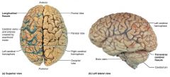
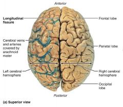
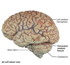
cerebrum |
brain region consisting of two cerebral hemispheres; 83% of total brain mass |
|
|
|
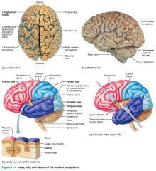
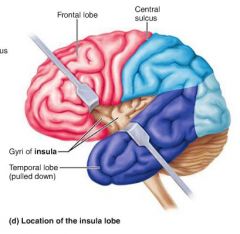
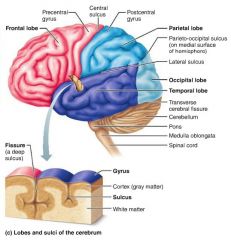
1. frontal 2. parietal 3. temporal 4. occipital 5. insula |
5 lobes of the cerebrum |

|
|
|
insula |
cerebral lobe buried deep to the lateral sulcus |
|
|
|
fissures |
deep grooves separating large regions of the brain |
|
|
|
1. longitudinal fissure 2. transverse cerebral fissure |
2 main brain fissures |

|
|
|
longitudinal fissure |
fissure separating the cerebral hemispheres |
|
|
|
transverse fissure |
fissure separating the cerebral hemispheres from the cerebellum below |
|
|
|
sulci |
shallow grooves in brain tissue |

|
|
|
gyri |
elevated ridges of brain tissue |
|
|
|
central sulcus |
groove separating the frontal lobe from the parietal lobe |

|
|
|
1. precentral gyrus 2. postcentral gyrus |
2 ridges bordering the central sulcus |

|
|
|
parieto-occipital sulcus |
groove separating the occipital lobe from the parietal lobe |

|
|
|
lateral sulcus |
deep groove separating the temporal lobe from the parietal and frontal lobes |

|
|
|
1. cerebral cortex 2. white matter 3. basal nuclei |
3 layers of a cerebral hemisphere (superficial to deep) |
|
|
|
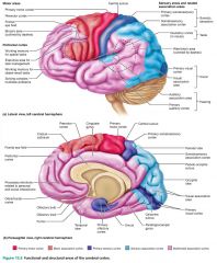
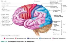
cerebral cortex |
the "executive suite" of the nervous system; enables self-awareness, communication, memory, understanding, and voluntary movement; 40% of total brain mass |
|
|
|
1. motor areas (anterior) 2. sensory areas (posterior) |
2 main functional areas of the cerebral cortex |
|
|
|
motor areas |
areas of the cerebral cortex that control voluntary movement; anterior to the central sulcus |

|
|
|
1. primary motor cortex 2. premotor cortex 3. Broca's area 4. frontal eye field |
4 motor areas of the cerebral cortex |

|
|
|
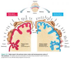
primary (somatic) motor cortex |
motor area that allows for skeletal muscle control and movement |

|
|
|
premotor cortex |
motor area that helps plan and coordinate more complex movements; ex. playing the piano |

|
|
|
Broca's area |
motor area that directs the muscles involved in producing speech; located on the left temporal lobe |

|
|
|
frontal eye field |
motor area that controls voluntary eye movement; anterior to premotor area |

|
|
|
sensory areas |
areas of the cerebral cortex concerned with conscious awareness of sensation; posterior to the central sulcus |

|
|
|
1. primary somatosensory cortex 2. somatosensory association cortex 3. visual areas 4. auditory areas 5. vestibular (equilibrium) cortex 6. olfactory cortex 7. gustatory cortex 8. visceral sensory area |
8 sensory areas of the cerebral cortex |

|
|
|
primary somatosensory cortex |
sensory area that receives information from sensory receptors in the body and identifies the area being stimulated |

|
|
|
somatosensory association cortex |
sensory area that integrates sensory inputs from the primary somatosensory cortex; identifies objects by size, texture, etc. |

|
|
|
primary visual cortex |
sensory area that receives visual information from the retina of the eye; located on the occipital lobe |

|
|
|
visual association area |
sensory area that communicates with the primary visual cortex and uses past visual experiences to interpret visual stimuli |

|
|
|
primary auditory cortex |
sensory area that receives and interprets sound; located on the temporal lobe |

|
|
|
auditory association area |
sensory area that perceives and recognizes sound stimuli; stores memories of sounds |

|
|
|
vestibular (equilibrium) cortex |
sensory area responsible for conscious awareness of balance; located in the insular lobe |
|
|
|
olfactory cortex |
sensory area that interprets smell; also associated with the limbic system (emotions); located inside the temporal lobe |
|
|
|
gustatory cortex |
sensory area that perceives taste stimuli; located deep to the temporal lobe |

|
|
|
visceral sensory area |
sensory area involved in conscious perception of visceral sensations |

|
|
|
multimodal association areas |
complexly connected areas of the cerebral cortex that give meaning to sensory information received; sensations, thoughts, emotions |

|
|
|
1. anterior association area 2. posterior association area 3. limbic association area |
3 main multimodal association areas |
|
|
|
anterior association area (prefrontal cortex) |
multimodal association area involved with intellect, complex learning abilities (cognition), recall, and personality; develops slowly |

|
|
|
posterior association area |
multimodal association area that recognizes patterns and faces, localizing us and our surroundings, and binding different sensory inputs into a coherent whole |
|
|
|
Wernicke's area |
part of the posterior association area that interprets words; posterior to Broca's area, together they help understand language |

|
|
|
limbic association area |
multimodal association area that provides the emotional impact that makes a scene important to us; memories |
|
|
|
lateralization |
each brain hemisphere has abilities not completely shared by its partner; division of labor |
|
|
|
cerebral dominance |
when a brain hemisphere has greater control over language abilities, math, and logic; the left hemisphere in 90% of people |
|
|
|
left hemisphere |
brain hemisphere that usually has greater control over language, math, logic, and handwriting |
|
|
|
right hemisphere |
brain hemisphere that usually has greater control over visual-spatial skills, artistic skills, and intuition |
|
|
|
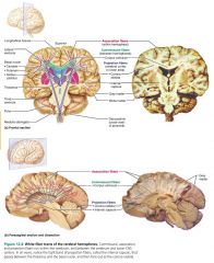
cerebral white matter |
layer deep to the cerebral cortex, consisting of myelinated fibers |

|
|
|
1. association fibers 2. commissural fibers 3. projection fibers |
3 types of fibers in cerebral white matter |

|
|
|
association fibers |
cerebral white matter fibers that connect different parts of the same hemisphere |

|
|
|
commissural fibers |
cerebral white matter fibers that connect corresponding areas of the two hemispheres |

|
|
|
corpus callosum |
largest commissural fiber connecting the left and right cerebral lobes |

|
|
|
projection fibers |
cerebral white matter fibers that connect to the lower brain and spinal cord |
|
|
|
internal capsules |
the bands of projection fibers at the top of each side of the brain stem |

|
|
|
corona radiata |
the fan-like arrangement of projection fibers, from the cerebral white matter to the cortex |

|
|
|
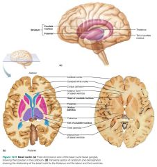
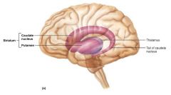
basal nuclei |
deepest cerebral region that helps control and monitor repetitive skeletal muscle movements |
|
|
|
1. caudate nucleus 2. putamen 3. globus pallidus |
3 main regions of the basal nuclei |
|
|
|
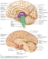
diencephalon |
brain region consisting of the thalamus, hypothalmus, and epithalamus; encloses the third ventricle |

|
|
|
1. thalamus 2. hypothalamus 3. epithalamus |
3 structures of the diencephalon |

|
|
|
thalamus |
structure of the diencephalon that serves as the gateway to the cerebral cortex; also involved in memory processing |

|
|
|
thalamic nuclei |
sort and receive sensory input from all over the body, and relay them to specific areas of the cerebral cortex; located in the thalamus |

|
|
|
hypothalamus |
structure of the diencephalon that maintains homeostasis; oversees the autonomic nervous system |

|
|
|
mammilary bodies |
hypothalamic nuclei that act as relay stations in the olfactory pathways |
|
|
|
infundibulum |
stalk of hypothalamic tissue connecting the pituitary gland to the base of the hypothalamus |

|
|
|
pituitary gland |
hypothalamic gland that secretes hormones |

|
|
|
epithalamus |
structure of the diencephalon that houses the pineal gland |

|
|
|
pineal gland |
epithalamic gland that secretes sleep-inducing hormone melatonin, which helps regulate the sleep-wake cycle |

|
|
|
brain stem |
"primitive" brain region that produces the automatic behaviors necessary for survival |

|
|
|
1. midbrain 2. pons 3. medulla oblongata |
3 regions of the brain stem |

|
|
|
midbrain |
region of the brain stem containing visual and auditory reflex centers; cranial nerves III and IV |

|
|
|
corpora quadrigemina |
four domelike nuclei that protrude dorsally from the midbrain; visual and auditory reflexes |

|
|
|
superior colliculi |
visual reflex nuclei of the midbrain that coordinate head and eye movements |

|
|
|
inferior colliculi |
auditory relay nuclei of the midbrain that relay sounds from hearing receptors to the sensory cortex |

|
|
|
1. substantia nigra 2. red nucleus |
2 motor nuclei of the midbrain |

|
|
|
pons |
brain stem region that relays information between the motor cortex and cerebellum; also helps with breathing; cranial nerves V through VII |

|
|
|
medulla oblongata |
brain stem region that receives instructions from the hypothalamus to control ANS functions; continuous with the spinal cord; cranial nerves VIII through XII |

|
|
|
pyramids |
two longitudinal ridges on the front of the medulla oblongata; corticospinal motor tracts |
|
|
|
decussation |
motor tracts of the medulla cross over before descending into the spinal cord |
|
|
|
visceral motor nuclei |
medullary nuclei that receive instructions from the hypothalamus |
|
|
|
1. cardiovascular center 2. respiratory center 3. various other centers |
3 functional groups of visceral motor nuclei in the medulla oblongata |
|
|
|
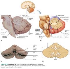
cerebellum |
brain region that subconsciously coordinates movements; ex. balance and posture |

|
|
|
vermis |
connects the right and left hemispheres of the cerebellum |
|
|
|
arbor vitae |
tree-like pattern of white matter branching through the cerebellum |

|
|
|
cerebellar peduncles |
motor tracts that connect the midbrain to the cerebellum; superior, middle, inferior |

|
|
|
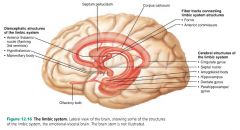
limbic system |
brain system that mediates emotional response; also involved in memory processing |

|
|
|
fornix |
fiber tracts linking limbic system regions together |
|
|
|
hippocampus |
structure of the limbic system responsible for long-term memory; along with the amygdaloid body |

|
|
|
psychosomatic illnesses |
emotion-induced illnesses |
|
|
|
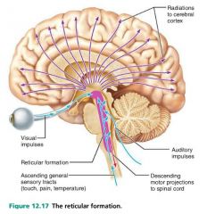
reticular formation |
brain system that keeps the cerebral cortex alert and active, and filters out repetitive stimuli; located in the brain stem |
|
|
|
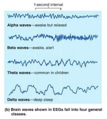
electroencephalogram (EEG) |
records electrical activity of neurons |
|
|
|
brain waves |
patterns of neuronal electrical activity measured by an EEG |

|
|
|
1. alpha 2. beta 3. theta 4. delta |
4 classes of brain waves |

|
|
|
alpha waves |
regular and rhythmic, low-amplitude, synchronous brain waves; awake but relaxed |

|
|
|
beta waves |
rhythmic, but less regular brain waves with a higher frequency; awake and alert |

|
|
|
theta waves |
irregular brain waves; more common in children or during concentration |

|
|
|
delta waves |
high-amplitude brain waves seen during deep sleep |
|
|
|
epileptic seizures |
seizures caused by a torrent of electrical discharges by groups of brain neurons |
|
|
|
petit mal (absence) |
mild seizures characterized by a blank expression and facial twitching; no loss of consciousness |
|
|
|
grand mal (tonic-clonic) |
severe seizures characterized by sensory hallucination, convulsions, and loss of consciousness |
|
|
|
consciousness |
conscious perception of sensations, voluntary movement, and higher mental processing |
|
|
|
1. alertness 2. drowsiness 3. sleep 4. stupor 5. coma 6. brain dead |
6 levels of consciousness |
|
|
|
fainting (syncope) |
a brief loss of consciousness |
|
|
|
coma |
significant unresponsiveness to sensory stimuli for an extended period; low oxygen use |
|
|
|
brain death |
a dead brain in an otherwise living body |
|
|
|
1. non-rapid eye movement (NREM) sleep 2. rapid eye movement (REM) sleep |
2 major types of sleep |
|
|
|
REM sleep |
stage of sleep characterized by increased oxygen use and heart rate, inhibited skeletal muscles, dreaming; alpha waves |
|
|
|
NREM sleep |
restorative stage of sleep, characterized by declining vital signs, possible nightmares or sleep walking |
|
|
|
slow-wave sleep |
deepest, restorative stages of NREM sleep (stages 3 and 4) |
|
|
|
circadian rhythm |
alternating cycle of sleep and wakefulness during a 24 hour period |
|
|
|
narcolepsy |
a condition characterized by the tendency to fall asleep abruptly |
|
|
|
insomnia |
a chronic inability to obtain the amount or quality of sleep needed to function adequately |
|
|
|
sleep apnea |
a temporary cessation of breathing during sleep |
|
|
|
1. Broca's area (speaking) 2. Wernicke's area (understanding) |
2 most important regions for language |
|
|
|
memory |
the storage and retrieval of information |
|
|
|
1. short-term memory (STM) 2. long-term memory (LTM) |
2 distinct stages of memory storage |
|
|
|
short-term memory (working memory) |
recent memory limited to seven or eight chunks of information; ex. phone number |
|
|
|
long-term memory |
memory with limitless capacity; storage and retrieval |
|
|
|
1. emotional state 2. rehearsal 3. association 4. automatic memory |
4 factors influencing the transfer of information from short-term memory to long-term memory |
|
|
|
memory consolidation |
the process of fitting new facts (memories) into categories of knowledge already stored in the cerebral cortex |
|
|
|
fact memory (declarative) |
memory that entails learning explicit information; ex. names, faces, words, dates |
|
|
|
skill memory (nondeclarative) |
memory that entails less conscious or unconscious learning; ex. playing a piano, riding a bike |
|
|
|
anterograde amnesia |
the loss of the ability to create new memories |
|
|
|
retrograde amnesia |
the loss of memories formed in the distant past |
|
|
|
1. skull 2. meninges 3. cerebrospinal fluid |
3 protective features of the brain |
|
|
|
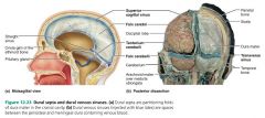
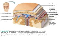
meninges |
connective tissue membranes that cover and protect the CNS, contain CSF, and form partitions in the skull |

|
|
|
1. dura mater 2. arachnoid mater 3. pia mater |
3 meninges of the brain |

|
|
|
dura mater |
two-layered, strong, external meninx that attaches to the skull |

|
|
|
dural sinuses |
spaces between the two dura mater layers of the brain that collect venous blood |

|
|
|
subdural space |
fluid-filled cavity between the dura mater and the arachnoid mater of the brain |

|
|
|
dural septa |
infoldings of the dura mater that partition areas of the brain |

|
|
|
1. falx cerebri 2. falx cerebelli 3. tentorium cerebelli |
3 dural septa of the brain |
|
|
|
arachnoid mater |
the loose, middle meninx |

|
|
|
subarachnoid space |
CSF-filled space full of web-like extensions that secure the arachnoid mater to the underlying pia mater |

|
|
|
arachnoid villi |
knob-like projections of the arachnoid mater that absorb CSF out of the subarachnoid space and into the superior sagittal sinus |

|
|
|
pia mater |
delicate, inner meninx that clings to the brain; full of tiny blood vessels |
|
|
|
meningitis |
inflammation of the meninges |
|
|
|
encephalitis |
inflammation of the brain |
|
|
|
cerebrospinal fluid (CSF) |
fluid that forms the liquid cushion in and around the brain |

|
|
|
choroid plexuses |
clusters of capillaries in the ventricles that produce cerebrospinal fluid |
|
|
|
hydrocephalus |
caused by CSF accumulation that exerts pressure on the brain |
|
|
|
blood brain barrier |
the protective mechanism that helps maintain the brain's stable environment |
|
|
|
endothelial cells |
form tight junctions that make brain capillaries the least permeable in the body; part of the blood brain barrier |
|
|
|
concussion |
an alteration in brain function following a blow to the head |
|
|
|
contusion |
bruising of the brain caused by serious concussions, can cause permanent neurological damage |
|
|
|
stroke (cerebrovascular accident) |
occurs when blood circulation to a brain area is blocked (ischemia) and brain tissue dies |
|
|
|
Alzheimer's disease (AD) |
a progressive generative disease of the brain ultimately resulting in dementia, memory loss, disorientation, etc. |
|
|
|
dementia |
mental deterioration |
|
|
|
Parkinson's disease |
results from a degeneration of dopamine-releasing neurons; characterized by persistent tremors, forward-walking posture and shuffling gait, and a stiff facial expression |
|
|
|
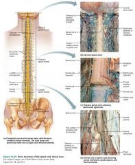
spinal cord |
two-way conduction pathway to and from the brain; also a major reflex center |

|
|
|
epidural space |
space filled with a soft padding of fat and a network of veins, between the vertebrae and the spinal dura mater |
|
|
|
1. vertebral column 2. cerebrospinal fluid 3. meninges |
3 protective features of the spinal cord |
|
|
|
spinal tap (lumbar puncture) |
procedure to remove CSF from below the spinal cord for testing (beyond L3) |
|
|
|
conus medullaris |
cone-shaped end of the spinal cord |
|
|
|
filum terminale |
fibrous extension of the conus medullaris that anchors the spinal cord to the coccyx |

|
|
|
deniculate ligaments |
saw-toothed shelves of pia mater that anchor the spinal cord to the vertebrae |

|
|
|
1. cervical enlargement 2. lumbar enlargement |
2 wide areas of the spinal cord that serve the upper and lower limbs |

|
|
|
cauda equina |
collection of nerve roots at the inferior end of the vertebral canal; "horse's tail" |

|
|
|
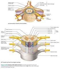
1. ventral median fissure 2. dorsal median sulcus |
2 grooves dividing the spinal cord into right and left halves |

|
|
|
central canal |
spinal canal containing cerebrospinal fluid |

|
|
|
gray commissure |
gray matter encircling the central canal of the spinal cord; butterfly-shaped |
|
|
|
ventral horn |
front column of motor neurons in the spinal cord |

|
|
|
dorsal horn |
rear column of sensory neurons in the spinal cord |

|
|
|
lateral horn |
lateral columns of sympathetic nerve fibers in the spinal cord |

|
|
|
white matter |
consists of nerve fibers that allow communication between the spinal cord and itself and the brain |
|
|
|
1. ascending 2. descending 3. transverse |
3 neuronal pathways connecting the brain and the body |
|
|
|
ascending pathways |
neuronal pathways that conduct sensory impulses upward to various areas of the brain |
|
|
|
1. first-order (receptor to spinal cord) 2. second-order (spinal cord to medulla) 3. third-order (medulla to cortex) |
3 sensory neurons of the ascending pathway to the brain |
|
|
|
sense ascending pathways |
neural pathways aware of sense but not location |
|
|
|
location ascending pathways |
neural pathways that locate the stimulus |
|
|
|
parallel pathways |
neural pathways with sense and location pathways ascending together, provide complete sensory information |
|
|
|
spinocerebellar tracts |
neural pathways that help coordinate skeletal muscle activity; run from proprioceptors to the spinal cord to the cerebellum |
|
|
|
descending pathways |
neuronal pathways that deliver efferent impulses from the brain to the spinal cord |
|
|
|
1. direct (pyramidal) 2. indirect (extrapyramidal) |
2 descending neuronal pathways from the brain |
|
|
|
1. planning 2. projecting 3. spinal |
3 levels of motor control (descending pathways) |
|
|
|
paralysis |
loss of motor function caused by damage to the spinal cord |
|
|
|
paraplegia |
loss of leg sensory and motor functions; spinal cord cut between T1 and L1 |
|
|
|
quadriplegia |
loss of sensory and motor functions below the head; spinal cord cut in the cervical region |
|
|
|
poliomyelitis |
viral disease that destroys ventral horn motor neurons; fever, headache, muscle pain, weakness, then paralysis |
|