![]()
![]()
![]()
Use LEFT and RIGHT arrow keys to navigate between flashcards;
Use UP and DOWN arrow keys to flip the card;
H to show hint;
A reads text to speech;
24 Cards in this Set
- Front
- Back
|
What are reflexes? |
- rapid, predictable motor responses to stimuli - mediated over neuronal pathways called Reflex arcs |
|
|
How can reflexes be functionally classified? |
1) Somatic reflexes - activate skeletal muscle 2) Autonomic (visceral) reflexes - activate visceral effectors (smooth or cardiac muscle or glands) |
|
|
What is reflex testing and how is it graded? |
It is an important diagnostic tool to assess the condition of the nervous system - reflex actions are graded according to responses from 0-4+ |
|
|
What are the components of a reflex arc? |
1) Receptor 2) Sensory Neurons 3) Integration center 4) Motor neurons 5) Effector |
|
|
What are the types of reflexes? (depending on number of neurons) |
a) Monosynaptic - 2 neurons involved
b) Polysynaptic - more than 2 neurons are involved (an association or interneuron is present) |
|
|
Diagram showing the 5 components of a reflex arc |

|
|
|
Where is the integration center for spinal somatic reflexes? Where are the effectors? |
Spinal somatic reflexes: Integration center - in spinal cord Effectors - skeletal muscle |
|
|
Why is it important to test the somatic reflexes? |
- allows assessment of the condition of the nervous system - if exaggerated, distorted, or asbent ==> degeneration/pathology of specific nervous system regions |
|
|
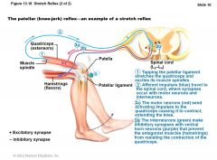
How does stretch reflex work? |
1) Stretch activates muscle spindle 2) Sensory neurons synapse directly w/ motor neurons in spinal cord 3) Motor neurons cause stretched muscle to contract |
|
|
True or false?
All stretch reflexes are monosynaptic and ipsilateral? |
TRUE
monosynaptic - only 2 neurons involved
ipsilateral - on the same side |
|
|
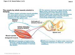
What is reciprocal inhibition of stretch reflexes? |
- fibers synapse w/ interneurons that inhibit motor neurons of antagonistic muscles ex) in patellar reflex, stretched muscle (quads) contracts and antagonists (hamstrings) relax |
|
|
Diagram explaining events by which muscle stretch is damped |
|
|
|
Diagram example of a patellar reflex |
|
|
|
What is a Flexor reflex? |
Flexor (withdrawal) Reflex 1) Initiated by PAINFUL stimulus 2) Causes automatic withdrawal of threatened body part 3) Ipsilateral and polysynaptic 4) Protective and important 5) Brain can override the reflex if needed ex) Finger stick for blood test |
|
|
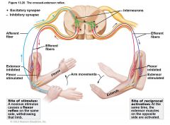
What is a Crossed-extensor reflex? |
1) Occurs w/ flexor reflexes in weight-bearing limbs to maintain balance
2) Consists of ipsilateral withdrawal reflex and contralateral extensor reflex
a) Stimulated side withdrawn (flexed) b) Contralateral side extended Ex) Step barefoot on broken glass |
|
|
diagram showing crossed-extensor reflex |

|
|
|
Another diagram showing a crossed-extensor reflex |
|
|
|
What are Superficial reflexes? |
- elicited by gentle cutaneous stimulation - depend on upper motor pathways and cord-level reflex arcs 1) Plantar reflex 2) Abdominal reflex (similar to when a MD is palpating the abdominal region, tense up) |
|
|
1) What areas of the spinal cord are tested through the Plantar reflex? 2) What stimulus would be used? 3) What response is expected? |
1) Tests integrity of cord from L4 - S2 2) Stimulus - Stroke lateral aspect of sole of foot 3) Response - Downward flexion of toes |
|
|
If there is damage to motor cortex or corticospinal tracts, what would occur when stimulating the Plantar reflex? |
Abormal response = Barbinski's sign - Hallux (big toe) dorsiflexes; other digits fan laterally - Normal in infants to ~1 year due to incomplete myelination |
|
|
1) What part of the cord does the Abdominal reflex test? 2) What is the stimulus and expected response? 3) Why would it be absent? |
1) Test integrity of cord from T8 - T12 2) Stimulus - stroking of skin Response - contraction of abdominal muscles and movement of umbilicus 3) Absent when corticospinal tract lesions are present |
|
|
What are the pupillary reflexes? |
Autonomic reflexes a) Pupillary light reflex b) Consensual reflex |
|
|
Which cranial nerves are tested with pupillary reflexes? |
1) Optic nerve CN II (is the receptor) 2) Occulomotor nerve CN III (conducts motor/efferent impulses to the smooth muscle of the iris which is the effector) |
|
|
What would absence of pupillary reflexes indicate? |
- a serious/severe trauma of vital brain stem issue |