![]()
![]()
![]()
Use LEFT and RIGHT arrow keys to navigate between flashcards;
Use UP and DOWN arrow keys to flip the card;
H to show hint;
A reads text to speech;
31 Cards in this Set
- Front
- Back
|
What does the Peripheral Nervous System do? |
- Gathers info from sensory receptors - Communicates w/ CNS and sends output to the effectors - includes neural structures outside the CNS: a) Sensory receptors b) Peripheral nerves and their ganglia c) Efferent motor endings |
|
|
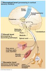
Organizations of PNS (diagram) |
|
|
|
What are sensory receptors classified by? |
- location - stimulus type |
|
|
What are the types of sensory receptors based on location? |
a) Exteroceptors: located on skin b) Interoceptors (visceroceptors) c) Proprioceptors |
|
|
What do Exteroceptors respond to? |
- respond to stimuli arising OUTSIDE body - touch, pressure, pain, temperature - most special sense organs |
|
|
What do Interoceptors respond to? |
- respond to stimuli arising in internal viscera and blood vessels - sensitive to chemical changes, tissue stretch, and temperature changes |
|
|
What do Proprioceptors respond to? |
- respond to stretch in skeletal muscles, tendons, joints, ligaments, and connective tissue coverings of bones and muscles - inform brain of one's movements |
|
|
What are the different sensory receptors based on stimulus type? |
a) Mechanoreceptors b) Thermoreceptors c) Photoreceptors d) Chemoreceptors e) Nociceptors |
|
|
What do Mechanoreceptors respond to? |
- respond to touch, pressure, vibration, and stretch |
|
|
What do Thermoreceptors respond to? |
- sensitive to changes in temperature |
|
|
What do Photoreceptors respond to? |
- respond to light energy |
|
|
What do Chemoreceptors respond to? |
- respond to chemicals |
|
|
What do Nociceptors respond to? |
- sensitive to pain-causing stimuli |
|
|
Diagram showing receptor level to perceptual level |
|
|
|
What is the structure of the nerve? Also, where do the different nerves arise? (Cranial, Spinal) |
Cordlike structures: consist of parallel bundles of axons enclosed in wrappings of connective tissue 1. Cranial nerves - arise from brain 2. Spinal nerves: arise from spinal cord |
|
|
Diagram showing nerve structure |
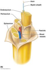
- note the epineurium, perineurium, and endoneurium |
|
|
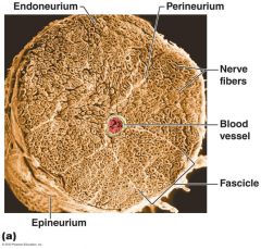
Picture showing nerve structure |
|
|
|
How are nerves classified? |
- most nerves are mixtures of Afferent and Efferent fibers and Somatic and Autonomic (Visceral) fibers
Classified according to direction of impulse: a) Mixed nerves - both sensory and motor; impulses to and from CNS b) Sensory (afferent) nerves - impulses towards CNS c) Motor (efferent) nerves - impulses only away from CNS |
|
|
Most nerves are mixed, what are the types of fibers in mixed nerves? |
a) Somatic afferent and Somatic efferent b) Visceral afferent and Visceral efferent |
|
|
What are Ganglia? |
Contain neuron cell bodies associated with nerves in PNS a) Ganglia associated w/ afferent nerve fibers contain cell bodies of sensory neurons (dorsal root ganglia - sensory, somatic) b) Ganglia associated w/ efferent nerve fibers contain autonomic motor neurons (Autonomic ganglia - motor, visceral) |
|
|
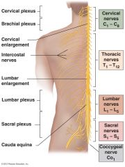
How many spinal nerves are there total, and how many in each section? |
31 pairs of mixed nerves named for point of issue from spinal cord
- 8 cervical (C1-C8) - 12 thoracic (T1-T12) - 5 Lumbar (L1-L5) - 5 Sacral (S1-S5) - 1 Coccygeal (C0) |
|
|
Diagram of the spinal nerves branching from the spine |
|
|
|
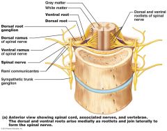
What are the root pairs that form the spinal nerves? |
1) Ventral roots: - contain motor (efferent) fibers from ventral horn motor neurons - fibers innervate skeletal muscles 2) Dorsal roots: - contain sensory (afferent) fibers from sensory neurons in dorsal root ganglia and conduct impulses from peripheral receptors |
|
|
Diagram showing the spinal nerve roots, ganglia, vertebrae, etc. |
|
|
|
How long are the spinal nerves? Also, what are the separate structures as they branch into mixed rami? |
Spinal nerve length: short (~1-2cm) Each branches into mixed rami: 1) Dorsal ramus 2) Ventral ramus - larger 3) Meningeal branch - tiny, re-enters vertebral canal, innervates meninges and blood vessels 4) Rami communicantes (autonomic pathways) join ventral rami in thoracic region |
|
|
Which ventral rami form interlacing nerve networks called nerve plexuses? |
- all ventral rami except T2-T12 form nerve plexuses |
|
|
How is the back innervated? |
- by dorsal rami via several branches |
|
|
What do the ventral rami of T2-T12 innervate? |
they become intercostal nerves: - supply muscles of ribs, anterolateral thorax, and abdominal wall |
|
|
Diagram showing a cross section of the thorax with the main roots and branches of a spinal nerve |

|
|
|
What is a dermatome? |
Dermatome - area of skin innervated by cutaneous branches of a single spinal nerve a) All spinal nerves except C1 participate in dermatomes b) Extent of spinal cord injuries ascertained by affected dermatomes c) Most dermatomes overlap, so destruction of a single spinal nerve will not cause complete numbness |
|
|
Multiple pictures not sure if necessary to add |
but we shall see |