![]()
![]()
![]()
Use LEFT and RIGHT arrow keys to navigate between flashcards;
Use UP and DOWN arrow keys to flip the card;
H to show hint;
A reads text to speech;
68 Cards in this Set
- Front
- Back
|
Functions of the Circulatory system |
1) transportation a. Respiratory gases, nutrients, and waste 2) regulation a. Hormonal and temperature i.e. water- ICF => interstitial fluid; temp => pH 3) protection a. Clotting and immunity I.e. infection, bleeding |
|
|
What's the job of cardiovascular system? |
Circulate the blood |
|
|
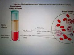
Blood |
Tissue type: connective epithelial tissue Consist of: Formed elements= WBC, RBC, PLT Plasma |
|
|
Cardiovascular system ( circulatory system) |
a. Heart: four-chambered pump b. Blood vessels: arteries, arterioles, capillaries, venules, and veins |
|
|
Lymphatic system (circulatory system) |
Lymphatic vessels, lymphoid tissues, lymphatic organs (spleen, thymus, tonsils, lymph nodes) |
|
|
Arterial blood |
Leaving the heart; bright red, oxygenated except for blood going to the lungs |
|
|
Venous blood |
Entering the heart; dark red deoxygenated except for blood coming from the lungs |
|
|
Constituents of blood |
|
|
|
Albumin (plasma proteins) |
Creates Osmotic pressure to help draw water from tissues into the capillaries to maintain blood volume and pressure. *big protein |
|
|
Globulins (plasma proteins) |
1) alpha and beta globulins - transport lipids and fat soluble vitamins 2) gamma globulins - antibodies that function in immunity. |
|
|
Fibrinogen (plasma proteins) |
Helps in clotting after becoming fibrin 1) serum - blood without fibrinogen |
|
|
Plasma volume |
a. Regulatory mechanisms maintain plasma volume to maintain blood pressure. b. Osmoreceptors in the hypothalamus cause the release of ADH from the posterior pituitary gland if fluid is lost. |
|
|
Erythrocytes RBC (formed elements of the blood) |
a. Flattened, biconcave discs b. Carry oxygen c. Lack nuclei and mitochondria d. Count - approx 5mil/mm blood e. Have 120 day lifespan f. Each contains about 280 million hemoglobin molecules g. Iron heme is recycled from the liver and spleen; carried by transferrin in the blood to the red bone marrow h. Anemia - abnormally low hemoglobin or RBC count |
|
|
Leukocytes (white blood cells- WBC) |
a. Have nuclei and mitochondria b. Diapedesis- movement through the capillary wall into connective tissue c. Count- approx 5k-9k/mm blood e. Types of leukocytes: granular, agranular |
|
|
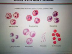
Types of leukocytes ( WBC) |
1) granular leukocytes: neutrophils, eosinophils, and basophils 2) agranular leukocytes whole in monocytes and lymphocytes |
|
Blood cells and platelets |
Formed elements |
|
|
Neutrophil (granulocytes) |
*multilobed nucleus (PMNL); inconspicuous cytoplasmic granules; diameter 10 to 12 * Phagocytize bacteria *Very phagocytic - "bacteria slayers" increase during acute bacterial infection and infection in general Neutrophilia: increased # of neutrophils Neutopenia: decreased # of neutrophils |
|
|
Eosinophil ( granulocytes) |
*Bilobed nucleus; red cytoplasmic granules; diameter 10-14 *Kill parasitic worms; complex role in allergy and asthma * Releases enzymes to digest parasitic worms Eosinophilia: increased # of eosinophils Eosinopenia: decreased # of eosinophils |
|
|
Basophil (granulocyte) |
* Dial of nucleus; large purplish black cytoplasmic granules; diameter 10-14 * Release histamine and other mediators of inflammation; contains Heparin, an anticoagulant *Rarest WBC *Basophilia: increase number of basophils *histamine: inflammatory chemical that acts as vasodilator to attract wbc's to inflamed sites *Are functionally similar to mast cells |
|
|
Lymphocytes ( agranulocytes) |
* spherical or indented nucleus; pale blue cytoplasm; diameter 5-17 *Mount immune response by direct cell attack or via antibodies *Act against virus-infected cells and tumor cells (cell mediated immunity) *B lymphocytes (b cell) give rise to plasma cells, which produce antibodies (antibody mediated immunity or humoral immunity) *Lymphocytosis: ⬆️ # of lymphocytes *Lymphopenia: ⬇️# of lymphocytes • increased in viral and fungal infection |
|
|
Monocyte (agranulocyte) |
* U or kidney-shaped nucleus; gray blue cytoplasm; diameter 14-24 * Phagocytosis; develop into macrophages in the tissues *Largest leukocyte *Highly phagocytic *the circulation enter tissues, and differentiate into macrophages * Monocytosis: increased monocytes |
|
|
Platelets (thrombocytes) |
a. Smallest form element, fragment of large cells called megakaryocytic b. Lack of nuclei c. Very short-lived (5- 9 days) d. Clot blood with several other chemicals and fibrinogen e. Release serotonin that stimulates vasoconstriction F. Count= 130k-49000 |
|
|
Haematopoiesis (hemopoiesis) |
1. Process of the blood cell formation a. Hematopoietic stem cells space embryonic cells that give rise to all blood cells b. Process occurs in myeloid tissue (red bone marrow) and lymphoid tissue c. As cells differentiate, they develop membrane receptors for chemical signals |
|
|
Erythropiesis |
a. Formation of red blood cells b. Red bone marrow produces about 2.5 million rbc's/sec c. Regulation of erythropoiesis 1) process stimulated by the way through erythropoietin from the kidneys that respond to low blood Erythopoietim evels 2) process takes about three days |
|
|
Erythropoiesis |
*Lose nucleus and become concave Stages=> [hemocytoblast (stem cell) -> Proerythroblast ->{Stimulated by erythropoietin} Erythroblast-> Normoblast->Reticulocyte ~ in bone bone marrow] =[Erythrocytes ~released into blood] |
|
|
Leukopoiesis |
a. Formation of white blood cells b. Cytokines stimulate the production of the different subtypes 1) multipotent growth factor-1 2) interleukin-1 3) interleukin-3 4) granulocyte colony-stimulating factor 5) granulocyte-monocyte colony-stimulating factor |
|
|
Antigens (RBC antigens and blood typing) |
Found on the surface of cells to help immune system recognize self cells |
|
|
Antibodies (RBC antigens and blood typing) |
Secreted by lymphocytes in response to foreign cells |
|
|
ABO system (RBC antigens and blood typing) |
Antigen or erythrocyte cell surface a. Type A has the A antigen b. Type B has the B antigen c. Type AB has both A and B antigens d. Type O has neither the A nor the B antigen |
|
|
Plasma contains antibodies against the antigens not present on the RBC |
Type A has anti B antibodies Type B has anti a antibodies Type A B has no antibodies (universal recipient) Type O has anti A and anti-B antibodies (universal donor) |
|
|
Transfusion reaction if a person receives the wrong blood type, antibodies bind to a Erythrocytes and cause ?? |
Agglutination |
|
|
Blood clotting |
1) hemostasis: cessation of bleeding when a blood vessel is damaged 2) damaged exposes collagen fibers to hold, producing: a.Vasoconstriction b. Formation of platelet plug c. Formation of fibrin protein web |
|
|
Mother Rh (-) & baby Rh (+) |
1st pregnancy(father Rh+) there is no issue because no antibody were made *Hemolytic anemia of newborn baby 2nd pregnancy (father Rh+) the mother has an antibody against baby |
|
|
How to prevent hemolytic anemia in newborn |
Giving mother rhogam |
|
|
Structure of the heart |
Four chambers Right atrium: receives deoxygenated blood from the body Left atrium: receives oxygenated blood from the lungs Right ventricle: pumps deoxygenated blood to the lungs Left ventricle pumps oxygenated blood to the body |
|
|
Fibrous skeleton (Structures of the heart) |
a. Separates Atria from ventricles. The Atria there for work as one unit, while the ventricles work as a separate unit. b. Forms that annuli fibrosi rings, which hold in heart valves |
|
|
Pulmonary (pulmonary and systemic circulations) |
Between heart and lungs Higher CO2 a. Blood pumps to lungs via pulmonary arteries b. Blood returns to heart via pulmonary veins |
|
|
Systemic (pulmonary and systemic circulations) |
Between heart and body tissues Higher O2 a. Blood pumps to body tissues via aorta. b. Blood returns to heart via Superior and inferior venae cavae |
|
|
Arteries |
Away from the heart |
|
|
Veins |
Back to the heart |
|
|
A/V atrioventricular valves |
Tricuspid=> right atrium and ventricle Bicuspid (mitral)=> left atrium and ventricle Papillary muscles and chordae tendineae prevent the valves from everting |
|
|
Semi-lunar valves |
Pulmonary: right ventricle and pulmonary trunk Aortic left ventricle and aorta |
|
|
Stethoscope positions for heart sounds (All Patient take medicine) |
Aortic area right Pulmonic area (nipple) left Tricuspid area left Bicuspid (Mitral) area right |
|
|
Cardiac cycle |
Repeating pattern of contraction and relaxation of the heart |
|
|
Systole (cardiac cycle) |
Contraction of heart muscles |
|
|
Diastole (cardiac cycle) |
Relaxation of heart muscles |
|
|
End-diastolic volume (cardiac cycle) |
Total volume of blood in the ventricles at the end of diastole |
|
|
End-systolic volume (cardiac cycle) |
The amount of blood left in the left ventricle after systole (1/3 of the end-diastolic volume) |
|
|
AV valves close during |
Systole |
|
|
AV valves open during |
Diastole |
|
|
Conduction systems of the heart |
Sa node => AV node =>Atrioventricle bundle ( bundle of his)=> purkinje fibers |
|
|
P wave |
Atrial depolarization |
|
|
PQ interval |
Atrial systole |
|
|
QRS wave |
Ventricular depolarization |
|
|
ST segment |
Plateau phase, ventricular systole |
|
|
T wave |
Ventricular repolarization |
|
|
Isovolumetric contraction (systole) |
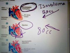
Volume of blood is the same |
|
|
Isovolumetric relaxation ( diastole) |
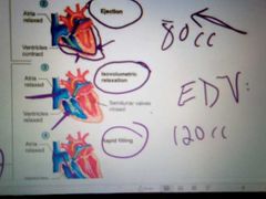
Same amount |
|
|
At the end of the diastole how much blood is there?? (EDV) |
120cc |
|
|
At the end of the systole how much blood is there?? (ESV) |
40cc |
|
|
What is the difference between EDV ESV is known as |
Stroke volume |
|
|
Incompetent valves (heart murmur) |
Do not close properly 1) maybe due to damage papillary muscle 2) mitral valve prolapse most common cause of chronic mitral regurgitation |
|
|
Septal defects (heart murmur) |
Holes in interventricular or interatrial septa which allows blood to cross the sides. |
|
|
Artery structure (tunica of blood vessels ) |
Tunica externa- outer layer; composed of connective tissue Tunica Media - middle layer => composed of smooth muscle for vasoconstriction/dialation Tunica interna =>inner layer; composed of simple squamous endothelium on a basement membrane and elastic fibers. |
|
|
Elastic arteries |
Aorta Closer to the heart; allow stretch as blood is pumped into them and recoil when ventricles relax |
|
|
Muscular arteries |
Farther from the heart; have more smooth muscle in proportion in diameter; also have more resistance do two smaller lumina |
|
|
Arterioles |
Smallest 20-30 um and diameter; provide the greatest resistance; control blood flow through the capillaries |
|
|
Capillaries (microcirculation) |
Smallest blood vessel Single layer of simple squamous epithelium tissue in wall Where are gases and nutrients are exchanged between the blood and tissues Blood flow to capillaries is regulated by a. Vasoconstriction and vasodilation of arterioles b. Precapillary sphincter
|