![]()
![]()
![]()
Use LEFT and RIGHT arrow keys to navigate between flashcards;
Use UP and DOWN arrow keys to flip the card;
H to show hint;
A reads text to speech;
36 Cards in this Set
- Front
- Back
|
Normal body temperature |
97.7° to 99.5° F (36.5° to 37.5° C) |
|
|
Pulse rate (adults) |
60-100 bpm |
|
|
Pulse rate (children) |
70-120 bpm |
|
|
Systolic blood pressure |
Occurs during cardiac contraction and should be less than 120mmHg |
|
|
Diastolic blood pressure |
Measured during relaxation of the heart and should be less than 80mmHg |
|
|
Normal respiration (adult) |
12-20 breaths/min |
|
|
Normal respiration (child) |
20-30 breaths/min |
|
|
Pulse oximeter |
Measures blood oxygen levels, which are normally between 95% and 100% |
|
|
Beta blockers |
May be used to reduce a patient's heart rate |
|
|
BUN and creatinine levels are used to: |
Indicate renal function |
|
|
Normal BUN |
7-25 mg/dL |
|
|
Normal creatinine |
0.5-1.5 mm/dL |
|
|
Normal BUN/creatinine ratio |
6:1 to 22:1 |
|
|
Normal GFR (men) |
70+/-14 mL/min/m^2 |
|
|
Normal GFR (women) |
60+/-10 mL/min/m^2 |
|
|
Prothrombin time; normal range |
Measure of blood coagulation; 12-15 seconds |
|
|
Coumadin/warfarin |
Anticoagulant |
|
|
Metformin/glucophage |
Used for treatment of type 2 diabetes; cease use X2 days following contrast administration |
|
|
_____ contrast agents are less likely to produce adverse side effects and/or reactions |
Non-ionic low-osmolar |
|
|
Routine transit time for contrast through the GI tract is: |
Between 30-90 minutes |
|
|
Examples of mild reactions to contrast |
N/V Mild urticaria (hives Pronounced sensation of warmth/flushing Alerted taste Sweats/chills Nasal stuffiness/sneezing Anxiety |
|
|
Examples of moderate reactions to contrast |
Mild bronchospasm Moderate to severe urticaria Vasovagal response Tachycardia from hypotension |
|
|
Examples of severe reactions to contrast |
Profound hypotension Laryngeal edema Severe bronchospasm Pulmonary edema Cardiac arrhythmia Seizure Cardiopulmonary arrest Death |
|
|
Factors affecting CT patient radiation dose |
System configuration User settings |
|
|
Slice sensitivity profile (SSP) |
Reconstructed CT section |
|
|
Dose profile |
Section of tissue exposed to ionizing radiation *greater than SSP |
|
|
Exposure; measured in _____ |
Ability of x-rays to ionized a volume of air; roentgens (R) |
|
|
Absorbed dose; measured in _____ |
Amount of x-ray energy absorbed in a unit of mass; grays (Gy) |
|
|
Kerma; air kerma |
Describes an absorbed dose; describes amount of radiation absorbed in a quantity of air |
|
|
Effective dose; measured in _____ |
Rush of exposure per certain types of tissue; sieverts (Sv) |
|
|
CT dose index (CTDI) |
Approx. Measure of the dose received in a single CT section or slice |
|
|
As the pitch increases, the dose per section (CTDIvol) _____ |
Decreases |
|
|
Pitch |
Amount of table travel per tube rotation divided by collimation |
|
|
Axial acquisition for brain |
Thin seconds (2-5mm)from skull base through posterior fossa, then 5-10mm sections through the vertex |
|
|
In brain scans, thinner sections through the posterior fossa are done to: |
Reduce beam-hardening artifact caused by the petrous pyramids |
|
|
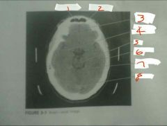
1. Frontal sinus 2. Falx cerebri 3. Right frontal lobe 4. Middle cerebral artery 5. Pons 6. Left temporal lobe 7. Quadrigeminal cistern 8. Cerebellum |