![]()
![]()
![]()
Use LEFT and RIGHT arrow keys to navigate between flashcards;
Use UP and DOWN arrow keys to flip the card;
H to show hint;
A reads text to speech;
86 Cards in this Set
- Front
- Back
|
Where is the heart located? |
Mediastinum |
|
|
What is the mediastinum? |
The area between the lungs, from the sternum to vertebral column. |
|
|
What is the apex of the heart? Where does it face? |
Blunt point of cone. Directed anteriorly, inferiorly, to the left. |
|
|
What is the base of the heart? Where does it face? |
Flace part opposite the apex. Directed posteriorly, superiorly, to the right. |
|
|
Where do the four points of the heart line up? |
Superior right: Superior border of 3rd costal cartilage Superior left: inferior border of 2nd costal cartilage. Inferior right: superior border of 6th costal cartialage Inferior left: 5th intercostal space Whole thing slightly shifted to the left. |
|
|
What is the outermost surface of the heart called? What two layers is it made up of? Describe them. |
Pericardium. Fibrous Pericardium - tough fibrous outer layer Serous Pericardium - thin transparent inner layer. |
|
|
What is the function of the fibrous pericardium? (2). |
Prevents over distention, acts as anchor. |
|
|
What is the serous pericardium made up of? |
Simple Squamous Epithelium |
|
|
What are the two layers of serous pericardium? |
Parietal Pericardium - lines fibour outer layer Visceral Pericardium - covers surface of heart |
|
|
What is the space between the two layers of the serous pericardium called? What fills it? |
Pericardial cavity. Pericardial fluid. |
|
|
What is another name for the visceral pericardium? |
Epicardium. |
|
|
What are the 3 layers of the heart wall? Briefly describe each |
Epicardium - visceral layer of serous pericardium Myocardium - cardiac muscle Endocardium - chamber lining and valves |
|
|
What are the four chambers of the heart? |
2 upper atria. 2 lower ventricles. |
|
|
What are Sulci? What do they contain (2)? |
Grooves on surface of heart. Contain coronary blood vessels and fat. |
|
|
What are the 3 sulci? What boundary does each mark? |
Coronary - marks boundary between atria and ventricles Anterior interventricular - boundary between ventricles (anteriorly) Posterior interventricular - boundary between ventricles (posterior) |
|
|
Which sulcus encircles the entire heart? |
Coronary. |
|
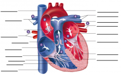
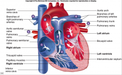
label dis bitch |
|
|
|
What are the two categories of valves in the heart? Describe each in terms of function. |
Atrioventircular - seperate atria and ventricles Semilunar - separate ventricles and arteries |
|
|
What are the two AV valves? |
Tricuspid valve - Right AV valve Bicuspid (mitral) valve - Left AV valve |
|
|
How do the semilunar valves function? |
Cusps fill up when pressure of blood in arteries is greater than pressure in heart, prevent blood from moving backwards into ventricles. |
|
|
Describe the pathway of coronary circulation. |
Aorta --> Coronary Arteries --> Heart Tissue --Coronary Sinus/Veins --> Right Atrium |
|
|
What are the anastomoses of the heart? What is their purpose. |
Redundant connections of the heart supplying blood to the same region. If one artery becomes occluded, alternate routes provide blood. |
|
|
Which artery supplies the posterior interventricular artery? The anterior? |
Right coronary artery supplies the posterior interventricular artery. Left coronary artery supplies the anterior interventricular artery. |
|
|
What are the two coronary veins? What do they drain? |
Great cardiac vein - left side of heart Small cardiac vein - right side of heart |
|
|
What is the coronary sinus? |
Large venous cavity that empties into the right atrium. |
|
|
What is the heart skeleton? What are its functions (2)? |
Fibrous plate (connective tissue) - allows for attachment of muscles and valves - insulates electrical signals to control APs |
|
|
How does the heart contract? |
Twists and pulls apex towards apex towards plate. "Wrings heart out." |
|
|
How many nuclei are in a cardiac muscle cell? Where is/are it/them located? |
1-2, centrally located |
|
|
Where are the t-tubules in cardiac muscle located? |
At the z-disc |
|
|
How big is the sarcoplasmic reticulum in cardiac muscle compared to skeletal muscle? What is it's location in terms of t-tubules? What implications does this have? |
Smaller comparatively. Doesn't contact t-tubules very often Causes increased contraction time |
|
|
How are cardiac muscle cells connected? |
Intercolated discs -specialized cardiac muscle cell-to-cell contacts. Many gap junctions. |
|
|
What intermembranous proteins connect cardiac muscle cells? Where are they found? |
Desmosomes (staples) and Gap Junctions Found at the intercalated discs. |
|
|
Where are the SA and AV Nodes located? |
SA: next to superior vena cava in right atrium AV: interatrial septum |
|
|
What is the function of the SA and AV nodes? |
Generate spontaneous action potentials. |
|
|
Which node is faster? What does this mean? |
SA. Is the one that actually starts the heart. |
|
|
What type of cells are the conducting system of the heart? How they vary in terms of structure and function from typical cells of the that type? |
Cardiac muscle cells. Less myofibrils - more for conducting than contracting. |
|
|
Where are APs conducted slowest in the heart? Why? |
AV node. Ensures the ventricles recieve AP after atria are done contracting. |
|
|
What is the pathway of conduction in the heart? |
SA Node --> Atria --> AV Node --> AV Bundle --> Left and Right Bundle Branches --> Purkinje Fibers |
|
|
Where is the fastest AP conduction in the heart? |
Purkinje Fibers > Left and Right Bundle Branches > AV Bundle |
|
|
How is AP conduction speed regulated in cardiac muscle cells? |
# of Gap Junctions |
|
|
Describe ion concentrations in cardiac muscle at rest. |
High extracellular Na+ and Ca2+, high intracellular K+ |
|
|
Describe cardiac muscle depolarization. |
Na+ channels open. K+ channels close Ca2+ channels slowly open |
|
|
What are the phases of repolarization in cardiac muscle action potential? |
Early repolarization. Plateau Phase Final Repolarization. |
|
|
What occurs in cardiac muscle early repolarization? |
Some voltage-gated K+ channels open. Na+ channels close. |
|
|
What occurs in cardiac muscle plateau phase? |
Some Ca2+ channels are open, slow repolarization. |
|
|
What is the effect of the plateau phase in cardiac muscle repolarization? |
Slows contraction. |
|
|
What occurs in Cardiac muscle final repolarization? |
Voltage-gated K+ channels open. Ca2+ channels close. Na/K ATPase restores ion gradient. |
|
|
What is the resting potential of cardiac muscle? |
-85 mV |
|
|
What happens if one cardiac muscle fiber fires? Why? |
They all fire. All APs are conducted from cell to cell. |
|
|
What is CICR? |
Calcium Induced Calcium Release. Movement of Ca2+ through plasma membrane and t-tubules into sarcoplasm stimulates release of Ca2+ from sarcoplasmic reticulum. |
|
|
What are the 3 phases of SA node AP? |
1) Pacemaker potential 2) Depolarization 3) Repolarization |
|
|
What occurs in the pacemaker potential phase of the SA node? |
Na+ leakage into the cell, causes resting potential to approach. K+ channels close (prevent repolarization) |
|
|
What occurs in the Depolarization phase of the SA node? |
Ca2+ channels open. K+ channels close.
|
|
|
What happens in the repolarization phase of the SA node? |
Ca2+ channels close, K+ channels open. |
|
|
How is sodium used in SA node AP? Calcium? |
Depolarization is almost entirely calcium. Sodium is mainly used to reach threshold. |
|
|
What are the two different refractory periods in cardiac muscle? Describe each. |
Absolute - completely insensitivity to stimulation Relative - reduced sensitivity to stimulation |
|
|
What is the point of long refractory periods in cardiac muscle? |
Prevent tetanic contraction. |
|
|
What are the 3 main phases of an ECG? |
P wave - atrial depolarization (contraction) QRS complex - ventricular depolarization and atrial repolarization T wave - atrial repolarization |
|
|
What happens in the PQ interval of an ECG? |
atrial contract and begin to relax |
|
|
What happens in the QT interval of an ECG? |
Ventricles contract and begin to relax. |
|
|
What are the two general states of the cardiac cycle? What do they refer to? |
Systole (Ventricular contraction) and Diastole (Relaxation) |
|
|
How many phases are there in Systole? What are they? |
2. Isovolumetric contraction and ejection |
|
|
How many phases are there in diastole? What are they? |
3. 1) Isovolumetric relaxation 2) Passive filling 3) Active ventricular filling |
|
|
When does active ventricular filling occur? |
During atrial systole. |
|
|
What happens in the following pressure situations? 1) Aortic > Atrial > Ventricular 2) Aortic > Ventricular > Atrial 3) Ventricular > Aortic > Atrial |
1) Semilunar valves closed, AV valves open, ventricular filling and atrial contraction 2) Semilunar valves closed, AV valves closed, isovolumetric ventricular contraction/relaxation or passive ventricular filling, atrial relaxation 3) Semilunar valves open, ventricular ejection, AV valves closed |
|
|
What is the equation for stroke volume? |
SV = End diastolic volume - end systolic volume |
|
|
What is the first heart sound? What does it coincide with? |
Lubb, AV valves closing at beginning of ventricular systole. |
|
|
What is the second heart sound? What does it coincide with? |
Dubb. Semilunar valves closing at beginning of ventricular diastole. |
|
|
When does a third heart sound occur? |
Turbulent flow into ventricles. |
|
|
What is the function of the chordae tendinae? |
Prevent eversion of the AV valves |
|
|
What is the function of the papillary muscles? |
Create tension in chordae tendinae |
|
|
What is the equation for mean arterial pressure? |
MAP = Cardiac Output x Peripheral Resistance |
|
|
What is the equation for cardiac ouput? |
Stroke Volume x Heart Rate |
|
|
What is the cardiac reserve volume? |
Difference between CO at rest and at maximum CO. |
|
|
How is the heart extrinsically regulated? |
PSNS and SNS |
|
|
What nerve and hormone are used by the SNS? What effect does this have?
|
Cardiac nerves = release of norepinephrine.
Increase HR (subsequently CO) Increase contraction force (Increase in SV, susbsequently CO) |
|
|
What nerve and hormone are used by the PSNS? What effect does this have? |
Vagus nerve = acetylcholine release Hyperpolarize heart, longer to reach threshold. (decreases HR) |
|
|
How does hormone control effect heart? |
(nor)epinephrine released from ardrenal medulla |
|
|
What two ways is the heart intrinsically regulated? |
Preload - amount of stretch controls force of contration Afterload - pressure the ventricle must produce to overcome pressure in aorta |
|
|
What is the law dealt with for preload? |
Starling's law of the heart |
|
|
At what speed does the release of ________ from the adrenal medulla effect the heart? |
Norepinephrine. Slowly, but for a longer period of time. |
|
|
How does body temperature affect heart rate? |
Positive correlation. |
|
|
How does extracellular ion concentration affect heart rate? |
Too much or too little extracellular K+ decreases HR. |
|
|
What portion of the brain senses pH and BP changes? |
Medulla Oblongata |
|
|
Where are chemoreceptors and baroreceptors found outside of the brain? |
Carotid and aortic bodies
|
|
|
Describe the baroreceptor reflex cycle for Blood Pressure. |
Low BP --> baroreceptors sense decreases --> cardioregulatory center in brain increases SNS and decreases PSNS --> (nor)epinephrine secretion from adrenal medulla --> increase HR and SV = increase in CO --> increase BP Opposite for high. |