![]()
![]()
![]()
Use LEFT and RIGHT arrow keys to navigate between flashcards;
Use UP and DOWN arrow keys to flip the card;
H to show hint;
A reads text to speech;
52 Cards in this Set
- Front
- Back
|
Longitudinal Section |
Lengthwise cut through any type of specimen |
|
|
Cross Section |
Crosswise cut through any type of specimen |
|
|
Metric System |
Standard system of measurement in the sciences. All conversions, whether for volume, mass (weight), or length are in units of ten |
|
|
Metric units of length |
Meter (m) Centimeter (cm) (10-2 m) Millimeter (mm) (10-3 m) Micrometer(um) (10-6 m) Nanometer (nm) (10-9 m) |
|
|
Metric units of weight |
Gram (g) = 1000mg Milligram (mg) |
|
|
Metric units of volume |
liter (L) = 1000mL millileter (mL) |
|
|
Metric Unit of Temperature |
Celsius (centigrade, C) C = (F-32)/1.8 F = (1.8C) + 32 |
|
|
Light Microscopes |
Passes rays of light through lenses to magnify the object |
|
|
Stereomicroscope or dissecting miscroscope |
Study entire object's surface in 3D at low magnification |
|
|
Compound light microscope |
Uses two sets of lenses (ocular and objective) and light to study an object Inverts image so that what is on the slide will appear backwards and upside down when viewed in the eyepiece Used for examining small or thinly sliced sections of objects under higher mangification than that of the steromicroscope |
|
|
Electron microscope |
- Uses beams of electrons to magnify the object. - The beams are focused on a photographic plate by means of electromagnets - Shows much more detail than a compound light microscope |
|
|
Resolution |
Minimum distance between two objects at which they can be seen or resolved as two separate objects (unit is __m) |
|
|
Parfocal nature of microscopes |
Being able to locate and focus your specimen at the lowest power objective before using a higher power objective and the object will be very nearly still in focus |
|
|
Total magnification |
Power of the objective lens * power of the ocular lens (unit is X) |
|
|
Stereomicroscope/Dissecting microscope and Compound Light microscope |
What are the 2 Types of Light Microscopes? |
|
|
Field of View |
Circle visible through the lenses |
|
|
Diameter of field |
Length of the field from one edge to the other |
|
|
Depth of field |
The area from top to bottom that comes into focus when viewing an object under high power |
|
|
Wet Mount |
A slide prepared by placing a drop of liquid or preparing a dry material with water or stain |
|
|
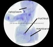
Cheek Cell: Identify cell membrane, cytoplasm, nucleus |
|
|
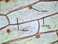
Onion Cell: Identify cell wall, cytoplasm, nucleus |
|
|
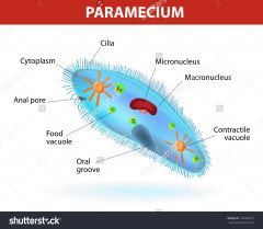
Paramecium: Identify cell membrane, cilia, oral groove, nucleus |
|
|
Cilia are important, because they are how Paramecium move. By beating the cilia back and forth, the Paramecium canmove through the water. |
How does a paramecium move? |
|
|
Compound |
Refers to the use of two sets of lenses: the ocular lenses located near they eyes and the objective lenses located near the object |
|
|
To improve contrast |
What is the purpose of using stains or dyes that bind to cellular structure and absorb light when examining specimens through a microscope |
|
|
Photomicrographs/Light micrographs |
Images produced by compound light microscope |
|
|
Transmission Electron Microscop |
- Analogous to the compound light microscope - Object is ultra-thinly sliced and treated with heavy metal salts to improve contrast |
|
|
Scanning electron microscope |
- Analogous to the dissecting light microscope - Gives an image of the surface and dimensions of an object (3D) |
|
|
Stereomicroscope (Dissecting) Scanning Electron Microscope |
Which two types of microscopes view the surface of an object? |
|
|
Compound Light microscope Transmission Electron Microscope |
Which two types of microscopes view objects that have been sliced and treated to improve contrast? |
|
|
Binocular head Eyepiece Lenses Focusing Knob Magnification Changing knob Illuminator |
Components of a Stereomicroscope (Dissecting) (5) |
|
|
Electron Microscopes |
What type of microscopes resolves the greater amount of detail? |
|
|
Binocular head |
Holds eyepiece lens(es) that move to accommodate for the various distances between different individuals' eyes |
|
|
Eyepiece lenses |
The lenses located on the binocular head |
|
|
Focusing Knob |
Large knob located on the arm; used for changing the focus of both eyepieces together |
|
|
Magnification changing knob |
Knob built into the binocular head, used to change magnification in both eyepieces simultaneously |
|
|
Illuminator |
Used to illuminate an object from above; may be built into the microscope or may be separate |
|
|
Eyepieces Viewing Head Arm Nosepiece Objectives Stage Mechanical Stage Adjustment Knobs Condenser Diaphragm control lever Light source Base |
Components of the Compound light microscope (12) |
|
|
Eyepieces |
Ocular Lenses |
|
|
Viewing head |
Holds the ocular lenses |
|
|
Arm |
Supports upper parts and provides carrying handle |
|
|
Nosepiece |
Revolving device that holds objectives |
|
|
Objectives |
- Scanning - Low Power - High power - Oil Immersion |
|
|
Stage |
Holds and supports microscope slides. |
|
|
Mechanical Stage |
Movable portion that aids in the accurate positioning of the slide |
|
|
Coarse adjustment knob |
Knob used to bring object into approximate focus; used only with low power objective |
|
|
Fine adjustment knob |
Knob used to bring object into final focus |
|
|
Condenser |
Gathers light from the lamp and directs it toward the object being viewed |
|
|
Diaphragm control lever |
Controls the amount of light passing through the condenser |
|
|
Light source |
Attached lamp that directs a beam of light up through the object |
|
|
Base |
Flat surface of the microscope that rests on the table |
|
|
- Optical axis of one corresponds exactly to the optical axises of the others
|
What does it means that the objectives of the microscope are concentric?
|