Reading...
![]()
Play button
![]()
Play button
![]()
Use LEFT and RIGHT arrow keys to navigate between flashcards;
Use UP and DOWN arrow keys to flip the card;
H to show hint;
A reads text to speech;
116 Cards in this Set
- Front
- Back
|
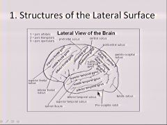
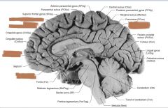
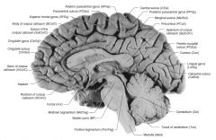
What sulcus seperates the parietal & frontal lobes from the limbic lobe?
|
Cingulate sulcus
|
|
|
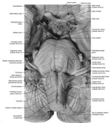
What cranial nerve exits through the interpeduncular fossa?
|

CN III
|
|
|
What cranial nerve exits dorsally and caudal to the inferior colliculi?
|

CN IV
|
|
|
Which one cranial nerve is best described as exiting to the lateral aspect of the basilar pons?
|

CN V
|
|
|
Which cranial nerve exits through the "ponto-medullary junction"?
|
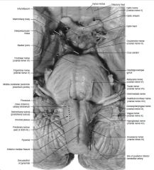
CN VI
|
|
|
Which cranial nerves (2) exit through the cerebellopontine angle?
|

CN VII & VIII
|
|
|
Which cranial nerves (3) exit through the "postolivary sulcus"?
|

CN IX, X & XI
|
|
|
Which cranial nerve exits through the "preolivary sulcus"?
|

CN XII
|
|
|
At which vertebral levels does the ventral horn enlarge laterally to accomodate autonomic function?
|
T1-L2 & S2-S4
|
|
|
What meningeal layer forms the denticulate ligaments?
|
Pia mater expands laterally to form multiple paired ligaments which attach to the arachnoid and dura mater.
|
|
|
At what vertebral level does the spinal cord end and what is the terminal portion called?
|
Conus medularis ends at L2
|
|
|
At what vertebral level does the dura and archnoid end?
|
S2
|
|
|
At what vertebral level does the pia mater end?
|
Filum terminale ends at the coccyx where it anchors the spinal cord in place
|
|
|
|
|
|
|
|
|
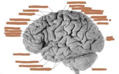
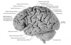
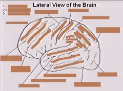
What gyrus is the "Primary motor cortex"?
|
Precentral gyrus
|
|
|
What gyrus is the "Primary somatosensory cortex"?
|
Postcentral gyrus
|
|
|
What two parts of the inferior temporal gyrus make up Broca's speech area?
|
Pars triangularis & Pars opercularis
|
|
|
What are the two subdivisions of the inferior parietal lobule?
|
Supramarginal gyrus & Angular gyrus
|
|
|
What is the Brodmann's area for Broca's motor speech area?
|
Left Hemisphere area 44 (pars opercularis) & 45 (pars triangularis)
|
|
|
What Brodmann's area is Wernicke's receptive speech area?
|
Left Hemisphere, posterior portion of area 22 (including planum temporale)
|
|
|
What Brodmann's area is the Primary motor cortex?
|
area 4 (precentral gyrus)
|
|
|
What Brodmann's area is the Primary somatosensory cortex?
|
area 3, 1, 2 (postcentral gyrus)
|
|
|
|
|
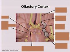
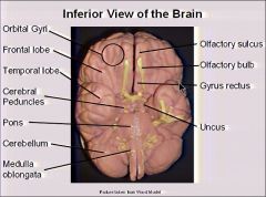
What does the Olfactory cortex consist of?
|
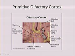
Olfactory bulb, Olfactory tract, Olfactory trigone, Piriform cortex, Periamygdaloid cortex & Entorhinal cortex
|
|

What does the Olfactory cortex consist of?
|

Olfactory bulb, Olfactory tract, Olfactory trigone, Piriform cortex, Periamygdaloid cortex & Entorhinal cortex
|
|
|
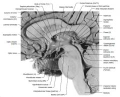
What is the "median hook" of the parahippocampal gyrus and where is it located?
|
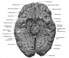
The "Uncus" is medial to the parahippocampal gyrus and lateral to the optic tract.
|
|
|
What is the Paracentral lobule?
|
It surrounds the medial aspect of the central sulcus and consists of the anterior & posterior paracentral gyrus.
|
|
|
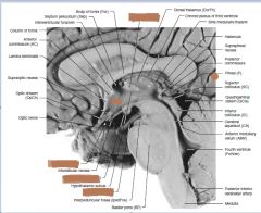
What two structures does the septum pellucidum seperate?
|
Lateral ventricles
|
|
|
What are the two things are seperated by the calcarine sulcus?
|
Cuneus & Lingual gyrus
|
|
|

|
|
|
Through what structure does the two occipital lobes communicate?
|
Splenium of corpus callosum
|
|
|
|
|
|
What is the CSF pathway from Lat. ventricles to the subarachnoid space?
|
Lat. vent.→Interventricular foramine (foramen of Monroe)→Third vent.→supraoptic recess of 3rd vent.→infundibular recess of 3rd vent.→cerebral aqueduct→fourth vent.→foramen of Luschka (lateral) & Magendie (medial)→subarachnoid space
|
|
|

|
|

|
|
|
|
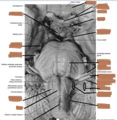
On a medial view, what are the landmarks for the Foramena of Luschka and Magendie?
|
Magendi - PICA (posterior inferior cerebellar artery
Luschka - Choroid plexus |
|
|
|
|
|
|
|
|
Define meninges?
|
Fibrous layers surrounding, protecting, and nourishing the CNS
|
|
|
Define venous sinus?
|
Large-diameter valveless venous vessel
|
|
|
Define cistern?
|
An expansion of the subarachnoid space resulting from irregularity of the surface of the brain, which is lined by pia mater
|
|
|
Define aneurysm?
|
Localized dilation of blood vessel, usually caused by atherosclerosis and hypertension, or, less frequently by trauma, infection or congenital weakness
|
|
|
What are the four cranial meningeal layers?
|
Dura mater - (periostialy & meningeal layer)
Arachnoids mater Pia mater |
|
|
What are two large septa of the brain formed by dural folds dwelling between the major subdivisions of the brain?
|
Falx cerebri & Tentorium cerebelli
|
|
|
Dural venous sinuses can be found where?
|
These large valveless vessels can be found between the two layers of dura mater
|
|
|
Which meningeal layer is avascular?
|
Arachnoid mater
|
|
|
Which meningeal layer is highly vascular?
|
Pia mater
|
|
|
What are the potential spaces within the cranium?
|
Subdural & epidural spaces
|
|
|
Between which two meningeal layers do the meningeal and cerebral arteries lie?
|
Meningeal - skull & dura
Cerebral - arachnoid & pia |
|
|
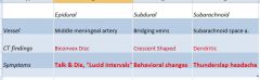
With regards to the three types of intracranial hemorrhages:
(3) Types? Common vessels involved? CT findings? Typical Symptoms? |
|
|
|
An Intracerebral hemorrhage reflects rupture of which arteries?
|
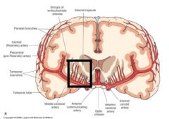
Intraparenchymal branches of subarachnoid arteries such as the lenticulostriate arteries branching from the middle cerebral artery and supplying the internal capsule and basal ganglia.
|
|
|
Which arteries are commonly injuried in obese people and infants resulting in an intracerebral hemorrhage?
|
Lenticulostriate arteries which branch from the middle cerebral artery.
|
|
|
|
|
|
Where does the right common carotid arise from?
|
brachiocephalic trunk
|
|
|
Where does the left common carotid arise from?
|
aortic arch
|
|
|
At what level do the common carotid arteries bifurcate?
|
They bifurcate at the level of the thyroid cartilage and become internal and external carotid.
|
|
|
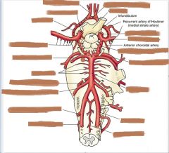
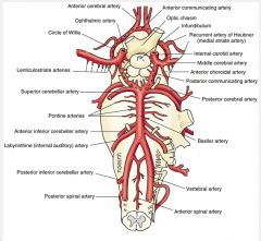
What four major cranial arteries arise from the internal carotid arteries?
|
Ophthalmic, posterior communicating arteries, middle cerebral & anterior cerebral
|
|
|
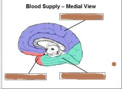
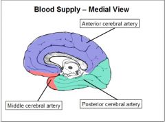
The medial surface of our brain is mainly supplied by what artery?
|
Anterior cerebral
|
|
|
|
|
|
|
|
|
Making the anterior cerebral arteries extremely important, what major structures on the medial aspect of the brain is fed by this artery?
|
Frontal & parietal lobes, anterior perforated substance, septum pellucidum and corpus callosum (often feeding all but the most posterior zones of this commisure)
|
|
|
What arteries supply the internal capsule and basal nuclei?
|
Lenticulostriate arteries
|
|
|
Making the middle cerebral arteries extremely important, which lateral lobes and (2) very important stuctures recieve blood via these arteries?
What are the specific arteries that supply the (2) important structures above? |
Frontal, temporal and parietal lobes...basal nuclei & internal capsule
Lenticulostriate arteries |
|
|

|
|
|
|
|
|
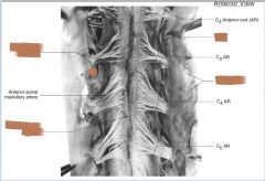
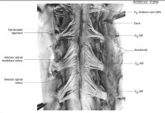
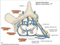
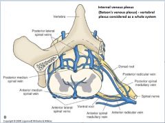
What are the (3) major arteries supplying the spinal cord?
What is the anastamotic plexus supplying the lateral portions and tied to the pia? |
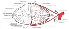
(2) posterior spinal arteries & (1) anterior spinal artery
The "arterial vasocorona" (AVC) is an anastamotic plexus supplying the lateral aspect with blood from the anterior and posterior spinal arteries. |
|
|
What are the major arterial contributors to the arterial vasocorona?
|

Segmental arteries that emit spinal medullary and redicular arteries
|
|
|
Brain arterioles constrict when systemic blood pressure is (lowered/raised) and dilate when systemic blood pressure is (lowered/raised) so that the cerebral blood flow is constant over what arterial pressure range?
|
Raised...Lowered
constant range of 60-150mm Hg |
|
|
When arterial CO2 is raised, how does the brain arterioles respond?
When the arterial O2 or pH are raised, how does the brain arterioles respond? |
Dilate and cerebral flow is increased
Constrict and cerebral flow is decreased |
|
|
What is epidural anesthesia?
|
Anesthetics injected into epidural space where they cause conduction block of adjacent spinal nerves
|
|
|
What is an aneurysm?
|
Dilation of the wall of an artery. Majority occur near branches of vessels (mostly internal carotid artery or its branches).
|
|
|
What is an embolism?
|
Results from an occlusion of a vessel by a clot, cells, gas. etc. This may interrupt blood supply, leading to tissue death.
|
|

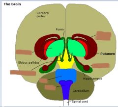
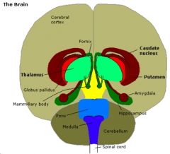
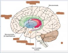
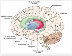
What is this structure?
|
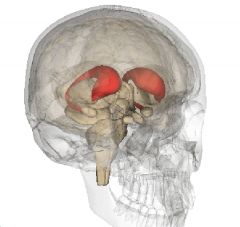
Caudate nucleus
|
|
Lateral Ventricles
|
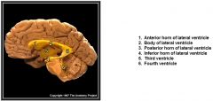
Lateral Ventricles
|
|
|
The floor and lateral walls of the lateral ventricles are formed by what important structure?
|
Caudate nucleus
|
|
|
What important structure forms the anterior wall of the inferior horn of lateral ventricle?
|
Amygdala
|
|
|
|
|
|
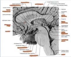
What forms the roof of the third ventricle? (2)
|
Choroid plexus and fornix
|
|
|
The lateral walls of the third ventricle is formed by what? (3)
|
Thalami, hypothalami & subthalami
|
|
|
What (2) structures form the anterior wall of the third ventricle?
|
Lamina terminalis & anterior commissure
|
|
|
What forms the posterior wall of the third ventricle?
|
Epithalamus
|
|
|
What (2) structures form the roof of the fourth ventricle?
|
Medullary vela & choroid plexus
|
|
|
What (2) structures denote the lateral boundries of the floor of the fourth ventricle?
|
Superior and inferior cerebellar peduncles
|
|
|
What (2) areas of the ventricular system do we not find choroid plexus?
|
Posterior (occipital) horn of lateral ventricles & cerebral aqueduct
|
|
|
What are the characteristics of CSF? (color, ions, cells)
|
CSF is a clear blood filtrate, low in glucose, protein, K+, Ca++ & cells
|
|
|
What are the (5) functional properties of CSF?
|
1)Bouyancy of brain
2)Shock absorption 3)Chemical stability 4)Reduction of Ischemia 5)Prevents traction of nerves |
|
|
What structures react to pressure gradients between the subarachnoid space and venous system, absorbing CSF through one way valves ensuring a unidirectional flow into the venous system?
|
Arachnoid granulations
|
|
|
What is a "non-communicating hydrocephalus" and what is another name for this condition?
|
An "obstructive (non-communicating) hydrocephalus results from obstruction of interventricular foramina, cerebral aqueduct or foramina of Luschka or Magendie and enlarging the undrained ventricles.
|
|
|
What is a "communicating hydrocephalus"?
|
Enlargement of all ventricles commonly reflective of impaired CSF absorption
|
|
|
|
|
|
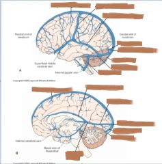
What are the (3) major sources of blood return to the cerebral sinuses?
|
1) Cerebral veins
2) Diploic veins 3) Emissary veins |
|
|
Where does the great anastomotic vein drain into?
|
superior sagittal sinus
|
|
|
Where does the small anastomotic vein drain into?
|
transverse sinus
|
|
|
The great cerebral vein (of Galen) forms at what junction of (2) veins and drains into what sinus?
|
The great cerebral vein (of Galen) forms at the two internal cerebral veins and drains into the straight sinus
|
|
|
The diploic veins are located where and communicate with which other veins?
|
Diploic veins lie between the layers of cranial bone, anastomose freely with each other and communicate w/ meningeal veins internally and superficial veins externally
|
|
|
Emissary veins connect which (2) other veins?
|
extracranial and intracranial veins
|
|
|
|
|
|
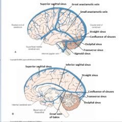
What are the (4) "unpaired dural venous sinuses" and what empties into each?
|
1) Superior longitudinal (sagittal) sinus - continuation of nasal vein receiving blood from cerebral, diploic & emissary veins
2) Inferior longitudinal (sgittal) sinus - receives blood from veins on medial surface of brain 3) Staight sinus - posterior continuation of great cerebral vein of Galen running between falx cerebri and tentorium cerebelli and receives blood from superior sagittal sinus and terminates at confluence of sinuses 4) Occipital sinus - receives blood from inferior cerebellar veins |
|
|
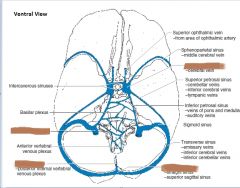
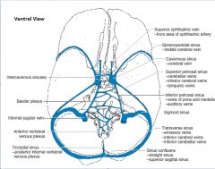
What are the (5) "paired dural venous sinuses" and what empties into each?
|
1) Transverse sinuses - receive blood from superior sagittal and straight sinuses and then draining into the jugular veins via sigmoid sinuses
2) Cavernous sinuses - (lateral to sphenoid bone) receives blood from superficial middle cerebral veins and superior ophthalmic veins (which communicate with each other via intercavernous sinuses, petrosal sinuses and pterygoid plexus of veins) ***Cavernous sinuses are largest and most important deep interconnecting sinuses which drain into internal jugular veins and transverse sinuses 3) Sigmoid sinuses - continuation of transverse sinuses receiving blood from the inferior cerebrum, cerebellum & emissary veins 4) Superior & inferior petrosal sinuses draining into transverse sinuses and internal jugular veins, respectively 5) Sphenoparietal sinuses - receive blood from superficial middle cerebral veins and drain into cavernous sinuses |
|
|
Where does the superior petrosal sinuses drain into?
|
Transverse sinuses
|
|
|
Where does the inferior petrosal sinuses drain into?
|
Internal jugular veins
|
|
|
|
|
|
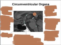
What is a circumventricular organ?
Name the (7) circumventricular organs? |
Organs positioned around the ventricular system, which have an incomplete blood-brain barrier resulting in these organs ability to directly sense concentrations of specific compounds
1) Pineal gland 2) Area postrema 3) Neurohypophysis 4) Organum vasculosum 5) Subfornical organ 6) Subcommissural organ 7) Median eminance of third ventricle |
|
|
|
|
|
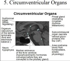
Name the (7) circumventricular organs and their function?
|
1) Pineal gland - mediate circadian rhythms (melatonin)
2) Area postrema - body fluid homeostasis, emetic physiology (induced vomiting) 3) Neurohypophysis - (post. pituitary) stimulates H2O retention and contraction of blood vessels to inc. BP 4) Organum vasculosum - fluid and electrolyte balance 5) Subfornical organ - thirst-regulating 6) Subcommissural organ - secrete various glycoproteins into CSF 7) Median eminence of third ventricle (floor) - hypophysiotropic hormones converge before B4 they are conveyed to the pituitary gland (neurohypophysis) |
|
|
What is a "Thrombosis of the dural sinus" due to?
|
Usually caused by a complication of middle ear, sinuses, nasopharynx, scalp or face infections.
Cavernous sinus thrombosis may be an unusual complication in diabetic patients, cortical phlebothrombosis may occur in non-infectious conditions such as hypercoagulable state following childbirth. |
|
|
During embryonic development, the Alar plate sits (dorsal/ventral) and the Basal plate sits (dorsal/ventral) to the sulcus limitans?
|
Alar plate sits dorsal and the basal plate sits ventral to the sulcus limitans.
|
|
|
What are the (3) major events tkaing place during formation of the mature brainstem?
|
1) Neural (central) canal enlarges into fourth ventricle
2) Cerebellum develops 3) Dorsal portion of neural tube (alar plate) rotates laterally Consequently many bulbar efferent structures lie medial to the sulcus limitans and afferent structures lie laterally. |
|
|
|
|
|
What makes up the "corpora quadrigemina"?
|
(2) each of superior and inferior colliculi
|
|
|
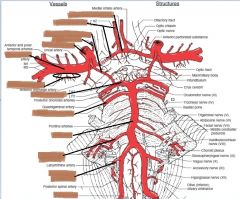
Where does the Pons get all of its blood supply?
|
Basilar artery
|
|
|
Where does the medulla get all of its blood supply?
|
PICA & anterior spinal artery
|
|
|
What does the labyrinthine artery supply?
|
Inner ear
|
|
|
|
|
|
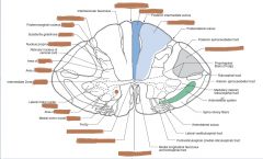
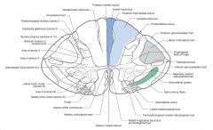
Wallenberg's (Lateral Medullary) Syndrome arises from occlusion of what artery?
What are the damgaed structures and correlated manifestations? |
PICA is occluded and results in damage to:
1) Spinal nucleus of trigeminal nerve - ipsilateral facial anesthesia/thermanesthesia 2) Anterolateral system - contralateral extrafacial anesthesia/thermanethesia 3) Inferior cerebellar peduncle - ipsilateral ataxias, uncoordination 4) Vestibular nuclei - vertigo & nystagmus 5) Efferents of CN IX & X - dysarthria & dysphagia 6) Disruption of hypothalamo-spinal fibers - Horners syndrome (ptosis, miosis, anhydrous) |