Reading...
![]()
Play button
![]()
Play button
![]()
Use LEFT and RIGHT arrow keys to navigate between flashcards;
Use UP and DOWN arrow keys to flip the card;
H to show hint;
A reads text to speech;
50 Cards in this Set
- Front
- Back
|
What kind of tissue encapsulates the kidneys?
|
Dense, irregular CT
|
|
|
What are the components of the renal cortex?
|
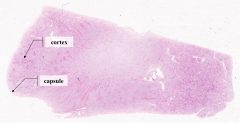
- Renal corpuscles
- Proximal and distal tubules - Capillaries - Medullary rays |
|
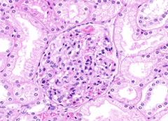
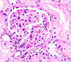
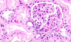
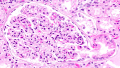
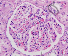
What is shown in this slide?
|

Renal Corpuscle:
- Glomerulus - Mesangial Cells - Bowman's Capsule |
|
What are Mesangial Cells? What is their function?
|
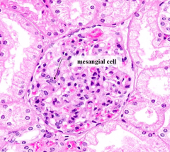
- Modified smooth muscle cells (pericytes)
- Support the glomerulus - May play a role in maintenance of the glomerular basement membrane |
|

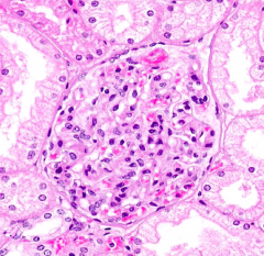
What structure can you see with the light microscope surrounding the glomerulus?
|

Parietal layer of Bowman's Capsule can be seen
|
|

What makes up the Parietal layer of Bowman's Capsule?
|
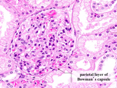
Simple Squamous Epithelium
|
|
|
What makes up the Visceral layer of Bowman's Capsule?
|
Podocytes
|
|
|
The nuclei within the glomerulus belong to what three types of cells? Can they be distinguished (if so, how)?
|
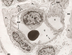
- Capillary Endothelial Cells (E)
- Podocytes (P) - Mesangial Cells - It is very difficult to distinguish them at the light microscope level w/o special staining techniques |
|
What is the clear space between the glomerulus and the parietal layer of Bowman's capsule called? What does this space contain?
|
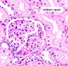
Urinary Space - contains a plasma ultrafiltrate (fluid that passed from the lumen of the capillaries into this space)
|
|
|
What components make up the filtration barrier through which fluid passes in going from the glomerular capillaries into the urinary space?
|
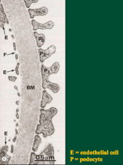
- Fenestrated Capillary Epithelium
- Fused Basal Laminae of capillary endothelial cells - Podocytes - Diaphragm-covered filtration slits between Podocyte foot processes |
|
What can be seen in this image?
|
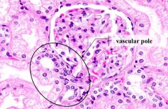
Vascular Pole
- Afferent arteriole supplies the glomerular capillaries - Efferent arteriole drains them |
|
What can be seen in this image?
|
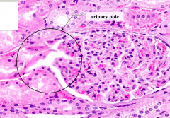
Urinary Pole
- Where plasma ultrafiltrate exits the renal corpuscle |
|
What can be seen in this image?
|
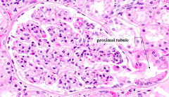
Proximal Tubule
- This is the first place the plasma ultriafiltrate enters into |
|
|
What is a nephron?
|
- Functional unit of the kidney
- Consists of a renal corpuscle, proximal tubule, loop of Henle, and distal tubule |
|
|
Proximal Tubules:
- Epithelium? - Lumen characteristics? - Cell borders? - Nuclei? - Cytoplasm? |

- High cuboidal
- Occluded lumen (d/t microvilli) - Indistinct cell borders - Few nuclei, basally located in a plane - Eosinophilic, granular cytoplasm |
|
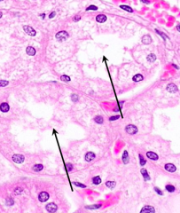
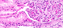
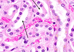
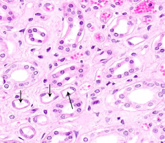
What kind of cells are these? How can you tell?
|

Proximal Tubule cells:
- High cuboidal - Occluded lumen (d/t long microvilli / brush border) - Indistinct cell borders (b/c lateral walls highly inter-digitated) - Few nuclei, basally located in a plane (b/c large cells) - Eosinophilic, granular cytoplasm (d/t lots of mito and basal membrane infoldings) |
|

What is the function of the cells in the Proximal Tubule?
|

- Passive reabsorption: Na+, Cl-, H2O
- Facilitated reabsorption: glucose, AA, proteins |
|
|
How does the number of Proximal Tubules compare to Distal Tubules in a given section? Why?
|
More Proximal Tubules / section because DTs are shorter than PTs
|
|
|
Distal Tubules:
- Epithelium? - Lumen characteristics? - Cell borders? - Nuclei? - Cytoplasm? |

- Low cuboidal epithelium
- Open, wide, smooth contouring lumen - Indistinct cell borders - Many, centrally located nuclei - Pale cytoplasm |
|

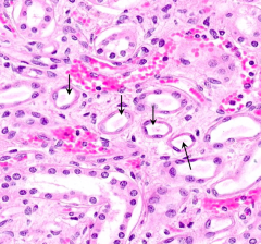
What kind of cells are these? How can you tell?
|
Distal Tubules:
- Low cuboidal epithelium - Open, wide, smooth contouring lumen (no brush border) - Indistinct cell borders (d/t extensive inter-digitations) - Many, centrally located nuclei (smaller cells, so more likely to have nuclei in section) - Pale cytoplasm |
|

What is the function of the cells in the Distal Tubule?
|

Resorption of Cl- and Na+
|
|

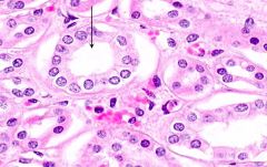
What kind of cells are circled? How can you tell?
|

Macula Densa of Distal Tubule
- Forms a disk of tightly packed columnar cells in the region of the vascular pole |
|
What is the function of the Macula Densa (circled)?
|

Monitors Na+ concentration and volume of the ultra-filtrate
|
|
|
What larger structure is the Macula Densa apart of that includes specialized cells in a portion of the afferent arteriole?
|
Juxtaglomerular Apparatus
|
|
|
What is the secretory product of the Juxtaglomerular Apparatus cells of the Afferent Arteriole?
|
Renin
|
|
|
In what part of the kidney do the thick descending limbs of the Loop of Henle descend?
|
Within Medullary Rays into the Medulla
|
|
|
Loop of Henle:
- Epithelium? |
- Thick part is cuboidal
- Thin part is squamous |
|
What kind of cells are these? How can you tell?
|
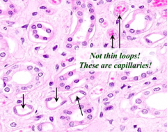
Thin part of Loop of Henle:
- Tubules lined by simple squamous epithelium, lacking RBCs (Tubules lined by simple squamous epithelium w/ RBCs in their lumens are capillaries) |
|
|
Collecting Duct:
- Epithelium? - Lumen characteristics? - Cell borders? - Nuclei? - Cytoplasm? |

- Cuboidal to Columnar epithelium
- Open, scalloped edge lumen because cells bulge inwards - Distinct cell borders - Many, centrally located nuclei - Pale cytoplasm |
|

What kind of cells are these? How can you tell?
|
Collecting Duct:
- Cuboidal to Columnar epithelium - Open, scalloped edge lumen because cells bulge inwards - Distinct cell borders - Many, centrally located nuclei - Pale cytoplasm |
|
|
What makes up the stroma of the kidney?
|
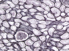
Primarily reticular fibers (although the stroma-supportive tissue is not as predominant as it is in other organs)
|
|
|
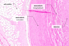
What are the layers/tunics of the ureter? Inner to outer?
|

- Mucosa - innermost
- Muscularis - middle - Adventitia - outermost |
|
|
What layer of the ureter supports the epithelium?
|
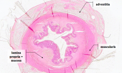
Lamina Propria / Mucosa
|
|
|
What type of epithelium lines the lumen of the ureter?
|
Transitional Epithelium
|
|

What is layer A?
|

Adventitia (outermost tunic) of ureter
|
|

What is layer B?
|

Muscularis (circular)
|
|

What is layer C?
|
Muscularis (longitudinal)
|
|
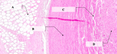
What is layer D?
|

Mucosa
|
|
|
How are the layers of the smooth muscle arranged in the muscularis layer of the ureter?
|

Upper 2/3 of ureter:
- Inner layer is longitudinal muscle - Outer layer is circular muscle |
|
|
How does the organization of the muscle in the muscularis layer of the ureter compare to the organization in the GI?
|

It is opposite, in the ureter:
- Inner layer is longitudinal muscle - Outer layer is circular muscle |
|
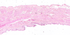
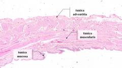
What are the layers/tunics of the bladder wall?
|

- Mucosa (inner)
- Muscularis (middle) - Adventitia (outer) |
|
|
What type of epithelium lines the lumen of the bladder?
|
Transitional Endothelium (Urothelium)
|
|
|
What are the components of the Tunica Mucosa in the Urinary Bladder?
|
- Transitional epithelium (urothelium)
- Underlying lamina propria (mostly fibrous CT) |
|
|
What kind of fibers are found within the lamina propria and muscularis of the bladder wall?
|
Elastin - refractile, pink staining fibers (they are much more coarse in the muscularis)
|
|
|
What layers of the bladder contain Elastin?
|
Lamina Propria and Muscularis (musch more coarse in Muscularis)
|
|

When fixed, this bladder was in what state?
|

Relaxed
|
|
|
Because the bladder is a saccular organ, how many layers does it have in the smooth muscle wall?
|
3
|
|
|
How common is urinary bladder cancer?
|
4th most common tumor in men and 8th most common tumor in women
|
|
|
What is a common presenting symptom of bladder cancer?
|
Gross, painless hematuria (90%)
|
|
|
Approximately 95% of bladder cancers arise from what cell type?
|
Transitional Cell (Urothelial) Carcinoma
|