Reading...
![]()
Play button
![]()
Play button
![]()
Use LEFT and RIGHT arrow keys to navigate between flashcards;
Use UP and DOWN arrow keys to flip the card;
H to show hint;
A reads text to speech;
43 Cards in this Set
- Front
- Back
|
What are the different methods for musculoskeletal imaging?
|
- Radiographs (plain films)
- Fluroscopy - Ultrasound - Computed Tomography (CT) - Magnetic Resonance Imaging (MRI) - Nuclear Medicine |
|
|
What things need to be considered when deciding on the appropriate imaging modality?
|
- Body part of interest
- Differential diagnosis consideration - Age of patient - Patient history - Cost of exam - Radiation dose - Availability |
|
|
What guidelines should be used to choose the appropriate imaging modality?
|
1. What is the clinical question?
2. What test is most likely to answer this question (what tests are available to you)? 3. If more than one test will work, consider safety and cost of the procedure |
|
|
How should you approach an imaging study?
|
- Develop a consistent search pattern
- E.g., hardware, joint(s), bones, soft tissues |
|

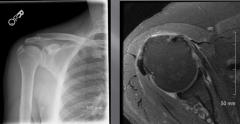
What is shown in this image?
|
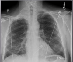
Pancoast tumor - lung apices don't look the same - tumor is causing shoulder pain
|
|
|
When you see one injury / fracture, what should your search pattern be next?
|
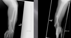
Look for another injury (often two)
E.g., Notice fractured radius, look at other angle to notice displaced ulna |
|
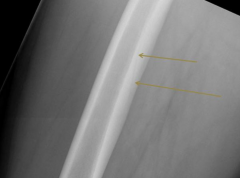
What is shown in this image?
|

Vascular Channel - not a fracture, tend to always be in same place so once you've seen them you should be able to recognize it again
|
|
|
What is a Sesamoid bone?
|
Bone embedded in a tendon where the tendon passes over a joint
|
|
|
What is the largest Sesamoid bone?
|
Patella
|
|
|
What is the name of the accessory bone that is occasionally seen in the posterior knee?
|
Fabella
|
|

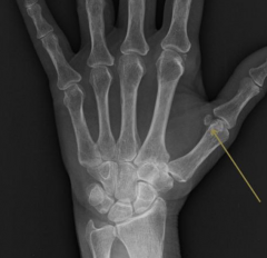
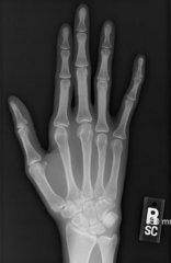
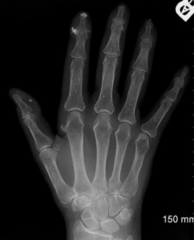
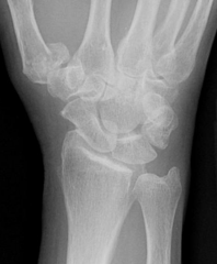
What is shown in this image?
|
Lunotriquetral Coalition - 7 instead of 8 wrist bones
(developmental abnormality) |
|

What is shown in this image?
|
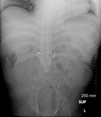
Sacral Agenesis (born without a sacrum)
|
|
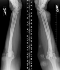
What is shown in this image?
|

Marfan's Syndrome (tall and lanky w/ long fingers)
|
|

What is shown in this image (look at the corners of the vertebrae)?
|
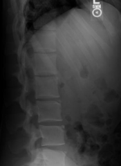
Limbus Vertebral Bodies (not a fracture, this is normal for them)
|
|
|
When looking at an image, what differential diagnoses should you consider?
|
VINDICATE:
- Vascular - Infection - Neoplasm - Drugs - Inflammatory / Idiopathic - Congenital - Autoimmune - Trauma - Endocrine / Metabolic |
|

What is shown in this image?
|
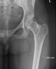
- Tooth in pelvis = Teratoma or got punched in face and swallowed tooth
- Fibrous Dysplasia (benign tumor) - caused lesion and a fracture through lesion |
|

What is shown in this image?
|
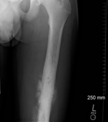
Osteosarcoma = malignant (not well defined)
|
|

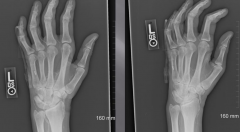
What is shown in these separate images?
|
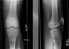
- Benign on left (more well-defined)
- Malignant on right (not well-defined) |
|
What is shown in this image?
|

Calcific Tendonitis
|
|
What is shown in this image?
|

Crest Syndrome - calcifications in soft tissue
|
|

What is shown in this image?
|
Soft Tissue Hemangiomas (abnormal calcification)
|
|
|
In what joints are effusions most easily diagnosed?
|
- Knee
- Elbow - Ankle - Wrist - Fingers |
|

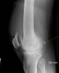
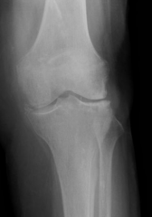
What is shown in this image?
|
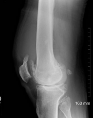
Effusion around knee (140 cc was removed)
|
|
|
How should you describe a fracture?
|
- Location
- Orientation - Displacement - Apposition - Angulation - Intraarticular involvement - Communication - Open vs. Closed - Associated injuries |
|
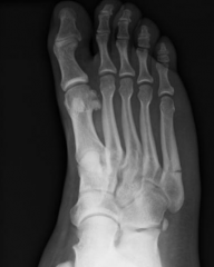
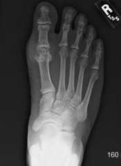
What is shown in this image?
|

Jones fracture (a fracture of the diaphysis of the fifth metatarsal of the foot)
|
|
What is shown in this image?
|

Rolando Fracture (intra-articular fracture through the base of the first metacarpal bone - the bone located just proximal to the thumb)
|
|

What is shown in this image?
|
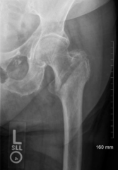
Intra-trochanteric Fracture - oblique fracture, very common, needs to be repaired
|
|
|
What happens in an avulsion fracture?
|
Fracture which causes the tendon or ligament to pull off of bone
|
|
|
What is the pattern on images for Osteoarthritis?
|
- Osteophytes
- Asymmetric joint loss |
|

What is shown in this image?
|
Gout
|
|
What is shown in this image?
|

CPPD (Calcium pyrophosphate dihydrate (CPPD) crystal deposition disease)
|
|
|
What are the most common images taken in radiology?
|
Radiographs (x-rays)
|
|
|
What are the pros and cons of Radiographs (xrays)?
|
Pros:
- Cheap - Readily available - Many different views and techniques - Often a good initial test Cons: - Radiation dose - Limited sensitivity - Does not work in all areas equally |
|
|
What is Fluoroscopy used for?
|
- Real time evaluation of structures
- Can be used intra-operatively - Used to set fractures and guid procedures - Interventional radiology, GI and GU radiology, lumbar punctures, joint injections, and biopsies |
|
|
What are the pros and cons of Fluoroscopy?
|
Pros:
- Real time evaluation of structures - Procedural guidance - Placement of needles, tubes, stents, and catheters Cons: - Radiation exposure - Image quality limited |
|
|
What is Ultrasound used for?
|
- Evaluate musculoskeletal system
- Guid procedures: biopsies, aspirations, and joint injections - Evaluate tendons and ligaments - Evaluate cortical surface of bone for erosions in diseases such as RA |
|
|
What are the pros and cons of Ultrasound?
|
Pros:
- No radiation - Portable machines / take up little space - No special room preparation Cons: - User dependent - Not all structures can be evaluated |
|
|
What is CT used for?
|
- Evaluate and characterize osseous lesions and fractures
- Evaluate superficial and deep infections - Evaluate post-traumatic and developmental deformities |
|
|
What are the pros and cons of CT?
|
Pros:
- Fast and available - Covers large anatomical area - Great for procedures Cons: - Over-utilization - Cost - Metal artifacts (from implanted devices) - Radiation dose |
|
|
What is MRI used for?
|
- Gold standard for evaluation of muscles, tendons, ligaments, and joints
- Evaluates cortical bone and marrow - Evaluate nearly every part of the body |
|
|
What are the pros and cons of MRI?
|
Pros:
- Great soft tissue detail - No radiation - Can obtain images in any plane Cons: - Expensive - Long exams - Metal artifacts - Claustrophobia |
|
|
What is Nuclear Medicine used for?
|
- Evaluates "physiology" and "function"
- Studies a radioactive material that is injected into patient; subsequent imaging shows distribution of material in body - A PET/CT scanner is a tool that combines radiology and Nuclear Medicine |
|
|
What are the pros and cons of Nuclear Medicine?
|
Pros:
- Functional data - Direct further radiology test - High sensitivity Cons: - Poor anatomical detail - Radiation dose - Low specificity - Expensive |